Clinical Hours: Mon-Fri, 8AM - 3PM(905) 493-5700
1885 Glenanna Road, Suite 220, Pickering, ON, L1V 6R6
An overview of skin cancer, from precancerous actinic keratosis to non-melanoma skin cancers and melanoma, with the surgical, topical, cryosurgical, and light-based treatments used to manage each.

Skin cancer is the most commonly diagnosed cancer worldwide, yet it is also among the most preventable and, when it is found early, the most treatable. The term covers a spectrum of conditions that ranges from precancerous lesions caused by cumulative sun exposure to invasive tumours capable of spreading to other organs. Understanding where a particular lesion sits on that spectrum determines how urgently it must be assessed and which treatment is most appropriate.
Most skin cancers arise in the epidermis, the outermost layer of the skin, and they are grouped by the cell type from which they originate. Keratinocyte cancers, historically called non-melanoma skin cancers, develop from the flat, keratin-producing cells that form the bulk of the epidermis. Melanoma develops from melanocytes, the pigment-producing cells scattered along the base of the epidermis. Between healthy skin and established cancer lies a category of precancerous change, of which actinic keratosis is the most common example.
The unifying cause across most of these conditions is ultraviolet radiation. Cumulative exposure from sunlight and indoor tanning damages the DNA of skin cells, and over many years that damage can overwhelm the repair mechanisms that normally keep cell growth in check. This shared origin explains why the same areas of sun-exposed skin, namely the face, ears, scalp, neck, forearms, and hands, tend to develop multiple lesions of different types over a lifetime.
An actinic keratosis is a rough, scaly patch that develops on chronically sun-damaged skin, and it can often be felt as a sandpaper-like roughness before it becomes clearly visible. Each individual lesion carries a low risk of progression, but that risk is not zero. A long-term analysis within the Veterans Affairs Topical Tretinoin Chemoprevention Trial found that many actinic keratoses regress on their own within a year, while a small proportion progress toward squamous cell carcinoma when they are left untreated [1].
Because actinic keratoses tend to appear in clusters across a broad region of sun-damaged skin, dermatologists describe the surrounding area as a field of cancerisation, meaning that it contains widespread microscopic damage beyond the visible spots. This concept matters for treatment, since addressing only the lesions that can be seen may leave subclinical abnormalities behind. Treatments that cover the entire affected field are therefore often preferred when the lesions are numerous.
Non-melanoma skin cancer, more accurately termed keratinocyte carcinoma, is by far the most common group of skin cancers. The two principal types, basal cell carcinoma and squamous cell carcinoma, differ in their behaviour, their appearance, and the urgency with which they must be treated, although both are strongly linked to ultraviolet exposure and both are usually curable when they are identified early.
Basal cell carcinoma is the most frequently diagnosed cancer in humans. It arises from the basal cells at the base of the epidermis and typically presents as a pearly or translucent bump, a non-healing sore, or a flat scaly patch, most often on the head and neck. It grows slowly and only very rarely spreads to distant sites, but when it is neglected it can invade and destroy the surrounding tissue, including cartilage and bone [2].
Squamous cell carcinoma is the second most common skin cancer and arises from the keratinocytes of the upper epidermis. It often appears as a firm, scaly or crusted nodule that may bleed or feel tender, frequently on the lips, ears, scalp, or backs of the hands. Unlike basal cell carcinoma, it carries a meaningful, although still modest, capacity to spread to the lymph nodes and beyond, which makes prompt treatment important [3].
Melanoma accounts for a minority of skin cancer diagnoses but the majority of skin cancer deaths, because it is able to spread to internal organs when it is not removed early. It develops from melanocytes and may arise within an existing mole or, more commonly, as a new pigmented spot. When melanoma is detected while it is still confined to the epidermis, it is almost always curable by surgery; once it has invaded more deeply, treatment becomes considerably more complex [4].
Early recognition rests on noticing change. The ABCDE guide summarises the features that help distinguish a concerning lesion from ordinary moles: asymmetry, border irregularity, colour variation, a diameter greater than six millimetres, and evolution over time. The evolving character of a lesion, whether in its size, shape, colour, or symptoms such as itching or bleeding, is regarded as one of the most reliable signals that a dermatological assessment is warranted [5].
Accurate diagnosis begins with careful examination of the skin, frequently supported by dermoscopy, a technique that uses a handheld magnifier with polarised light to reveal structures beneath the surface that are not visible to the naked eye. A periodic skin cancer screening examination allows suspicious lesions to be identified in a systematic way, which is particularly valuable for people with fair skin, extensive sun damage, numerous moles, or a personal or family history of skin cancer.
When a lesion appears suspicious, a definitive diagnosis requires a skin biopsy, in which a sample of tissue is removed and examined under the microscope by a pathologist. The biopsy confirms whether cancer is present, identifies its type, and establishes how deeply it extends. These findings guide the choice of treatment, because the optimal approach differs substantially between a superficial precancer and an invasive tumour.
Treatment is selected according to the type of cancer, its size, its location, its depth, and the general health of the patient. Superficial and precancerous lesions can often be managed with treatments applied to the surface of the skin, while invasive cancers are usually removed surgically. The main approaches are outlined below.
Surgery is the mainstay of treatment for most invasive skin cancers. A standard surgical excision removes the tumour together with a margin of normal-appearing skin, which is then checked under the microscope to confirm complete removal. For tumours in cosmetically or functionally sensitive areas, or those with aggressive features, Mohs micrographic surgery examines the tissue layer by layer during the procedure, sparing healthy skin while confirming clear margins, and skin cancer surgery of this kind achieves very high cure rates. Smaller superficial lesions may instead be treated with curettage and cautery, in which the abnormal tissue is scraped away and the base is sealed with heat.
Cryosurgery treats lesions by freezing them with liquid nitrogen, which destroys abnormal cells by forming ice crystals within them. It is a rapid, office-based option that is well suited to actinic keratoses and some superficial cancers, and it requires neither an incision nor stitches. A prospective study of cryosurgery for actinic keratosis reported that clearance rates improved with longer freeze times, which illustrates how technique influences the outcome [6]. Temporary blistering, redness, and a pale mark after healing are common.
Topical chemotherapy uses a cream that the patient applies at home to treat precancerous lesions and some very superficial cancers across an entire affected field. The most established agent is 5-fluorouracil, which interferes with DNA synthesis in rapidly dividing abnormal cells and produces a period of redness and inflammation as those cells are cleared. In a randomised trial that compared four field treatments for actinic keratosis, 5-fluorouracil cream was the most effective at keeping the treated area clear twelve months later [7]. These prescription creams are dispensed and monitored by the treating physician.
Topical immunotherapy works differently, by recruiting the body's own immune system rather than attacking the cells directly. Imiquimod, the most widely used agent, stimulates local immune receptors that provoke an inflammatory response against abnormal cells, and it is used for actinic keratoses and for superficial basal cell carcinoma. The same randomised comparison of field treatments found imiquimod to be effective as well, although a course requires several weeks of application and a predictable inflammatory reaction in the treated skin [7].
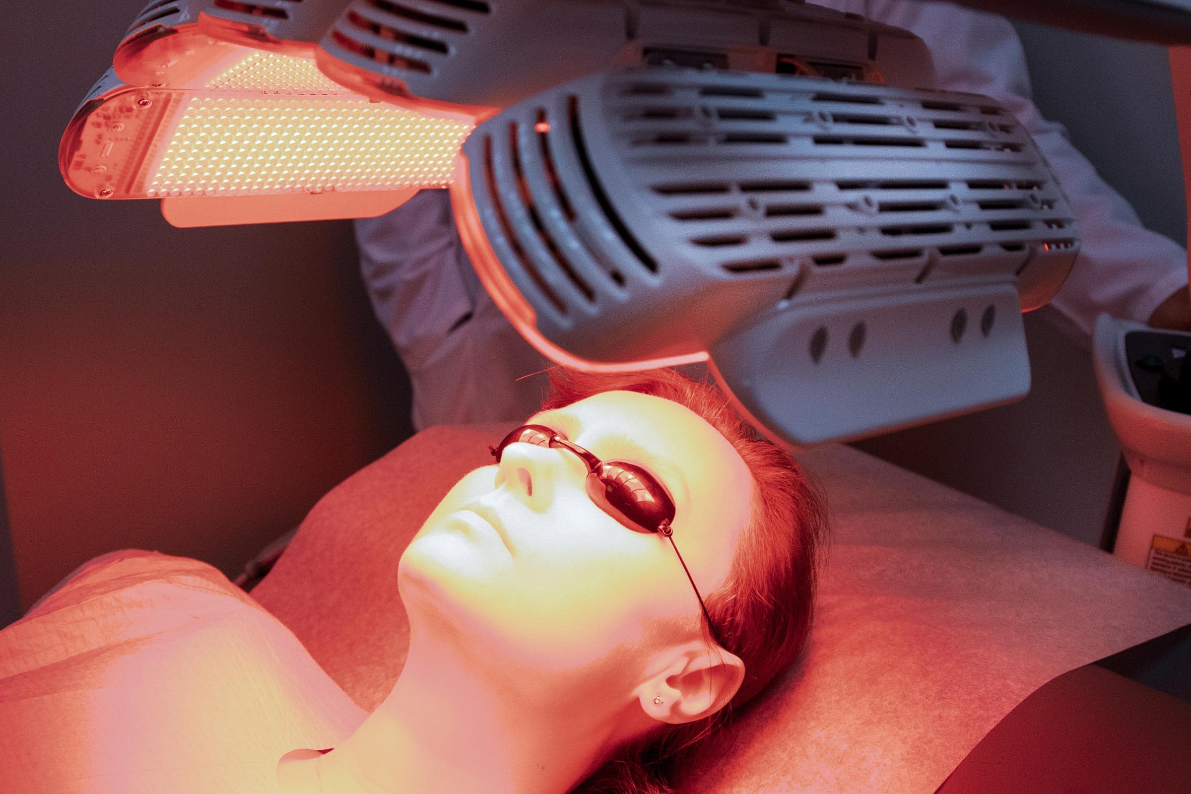
Photodynamic therapy combines a light-sensitising cream with a specific wavelength of light. The cream is preferentially absorbed by abnormal cells and, once it is activated by the light, generates reactive oxygen that destroys those cells while largely sparing the surrounding healthy skin. It is especially useful for treating multiple actinic keratoses across broad areas such as the scalp or face, and for selected superficial cancers, with excellent cosmetic results. The mechanism behind this treatment, and its surprising connection to historical vampire legends, is explored in a related article on photodynamic therapy.
Because ultraviolet radiation drives the great majority of skin cancers, prevention centres on reducing exposure. Consistent sun protection, including broad-spectrum sunscreen, protective clothing, hats, and shade during peak hours, measurably lowers the risk of both keratinocyte cancers and melanoma, and it slows the development of new actinic keratoses in skin that is already damaged. Avoiding indoor tanning is equally important, because artificial ultraviolet sources carry the same risks as the sun.
Early detection is the other half of the strategy. Regular skin self-examination, with attention to any lesion that is new, changing, or failing to heal, helps bring suspicious spots to attention sooner. For anyone who has already been treated for a skin cancer or a precancer, structured post-treatment surveillance is recommended, because sun-damaged skin remains at elevated risk of developing further lesions long after the original one has been treated.
Any spot that changes in size, shape, or colour, that bleeds without healing, or that simply looks different from the skin around it deserves professional evaluation. A consultation with a dermatologist allows suspicious lesions to be examined, biopsied when necessary, and treated with the approach best suited to the diagnosis, which offers the best chance of a straightforward cure.
This article is intended for educational purposes and does not replace professional medical advice. Please consult your dermatologist for personalized recommendations.
Your feedback helps us improve our news and clinical insights
From medical dermatology to surgical procedures, our clinic provides comprehensive care for all skin, hair, and nail conditions.