Clinical Hours: Mon-Fri, 8AM - 3PM(905) 493-5700
1885 Glenanna Road, Suite 220, Pickering, ON, L1V 6R6
Solar lentigines, melasma, post-inflammatory pigment, and lentigo maligna can all look alike. How brown facial spots are told apart, and what IPL, lasers, and peels realistically achieve.

A brown spot on the face is a description, not a diagnosis. The same flat tan macule on a cheek may be a solar lentigo, a patch of melasma, the residue of an inflammatory lesion that healed months earlier, an early seborrheic keratosis, or, less commonly, a lentigo maligna. Each of these arises from a different process, sits at a different depth within the skin, and responds differently to treatment. Choosing a laser or a peel before the lesion has been identified is the most common error in the management of facial pigmentation.
Solar lentigines are flat, sharply bordered tan to dark brown macules that accumulate on chronically sun-exposed skin from roughly the fourth decade onward. They reflect a localised increase in melanocyte number and activity, with melanin accumulating within the epidermis. Ephelides, commonly called freckles, are smaller, appear in childhood in fair-skinned individuals, and darken in summer before fading in winter. Both are frequently present on the same face, and both are epidermal, which is what makes them the most treatable form of facial pigmentation [1].
Melasma presents as symmetric brown to grey-brown patches across the cheeks, forehead, upper lip, and jawline, most often in women with intermediate skin phototypes. It is not simply pigment sitting in the epidermis. Current models describe a damaged basement membrane, senescent dermal fibroblasts, increased vascularity, and melanin that has descended into the dermis, which together explain why melasma relapses so readily once treatment is stopped [2]. Ultraviolet radiation, visible light, and hormonal influence including pregnancy and combined oral contraceptives all act as drivers.
Post-inflammatory hyperpigmentation is pigment left behind after inflammation has resolved. Acne, dermatitis, and cosmetic procedures themselves are the usual antecedents. The pigment may be epidermal, in which case it fades over months, or dermal, in which case it can persist for years. It is the single most important complication to anticipate when any device or peel is applied to darker skin, because aggressive treatment of pigmentation can generate more pigmentation.
A pigmented seborrheic keratosis is a benign overgrowth of keratinocytes rather than a collection of melanin, even though it can be uniformly brown. The surface is waxy or warty and the border stops abruptly against normal skin, giving the impression that the lesion has been placed onto the surface rather than grown within it. Pigment-selective lasers are the wrong instrument for these lesions, which are removed by curettage or cryosurgery when removal is warranted.
Lentigo maligna is a melanoma in situ that arises on chronically sun-damaged skin of the head and neck. It presents as a slowly enlarging macule with irregular pigmentation and an indistinct border, and it is mistaken for a solar lentigo often enough that this confusion defines the clinical problem. Its prolonged radial growth phase means that a lesion may be present for years before invasion occurs, and that same indolence is what allows it to be dismissed as a harmless age spot [3].
The consequence of getting this wrong is not simply a treatment failure. A review of patients who presented with melanoma after previous laser treatment of a pigmented lesion documented how destruction of a lesion that was never examined histologically removes the evidence, delays the correct diagnosis, and complicates staging when the melanoma later becomes apparent [4]. No tissue remains for the laboratory once a lesion has been vaporised or shattered.
The rule that follows is absolute. A pigmented facial lesion is assessed before it is treated, not after. Dermoscopy resolves most of the uncertainty by revealing subsurface structures that clinical inspection cannot show, and a skin biopsy settles the remainder. Only once malignancy has been excluded does the question of which device to use become reasonable.
Every pigment-directed treatment operates on the principle of selective photothermolysis: energy is delivered at a wavelength that the target absorbs more strongly than the surrounding tissue, and in a pulse short enough that the heat generated does not spread beyond the target before it dissipates [5]. Melanin absorbs light across a broad range of wavelengths, with absorption falling progressively as wavelength increases. Shorter wavelengths are therefore strongly absorbed but penetrate only superficially, while longer wavelengths reach deeper but with less selectivity.
This physics explains the clinical pattern. Epidermal pigment, as found in solar lentigines and ephelides, is close to the surface and highly accessible, which is why these lesions often clear in one or two sessions. Dermal pigment requires longer wavelengths, more sessions, and greater caution. Melasma, which is mixed and continuously re-driven by light and hormonal factors, is the least device-responsive of the common diagnoses despite being the one patients most often ask to have lasered.
Photoprotection is not an adjunct to the treatment of facial pigmentation. It is the treatment on which everything else depends. Broad-spectrum sunscreen at SPF 50 or higher, applied daily and reapplied through the day, both prevents new solar lentigines from forming and prevents treated ones from returning.
Ultraviolet protection alone is incomplete for melanin-rich skin. Visible light has been shown to induce pigmentation in darker phototypes that is both darker and more sustained than that produced by equivalent doses of ultraviolet A [6]. Conventional sunscreens are transparent to visible light, so tinted formulations containing iron oxides are required. In a randomised comparative trial, a sunscreen protecting against short wavelengths of visible light reduced melasma relapse relative to a sunscreen offering ultraviolet protection alone [7].
Topical therapy remains first line for melasma and is a reasonable starting point for lentigines. The fixed triple combination of hydroquinone 4 percent, tretinoin 0.05 percent, and fluocinolone acetonide 0.01 percent outperformed hydroquinone alone in a randomised controlled trial in patients with moderate to severe melasma [8]. A Cochrane review of interventions for melasma found the triple combination to be the best supported topical option while noting that the overall evidence base is limited by small trials, short follow-up, and heterogeneous outcome measures [9].
Two further options carry useful evidence. Oral tranexamic acid produced significantly greater improvement than placebo in a randomised, double-blind trial in moderate to severe melasma, with the benefit diminishing after the drug was stopped [10]. Topical cysteamine 5 percent, which does not carry the risk of exogenous ochronosis associated with prolonged hydroquinone use, outperformed placebo in a randomised double-blind trial in epidermal melasma [11]. Azelaic acid, topical retinoids, and niacinamide are used as adjuncts and as maintenance once an initial response has been achieved.
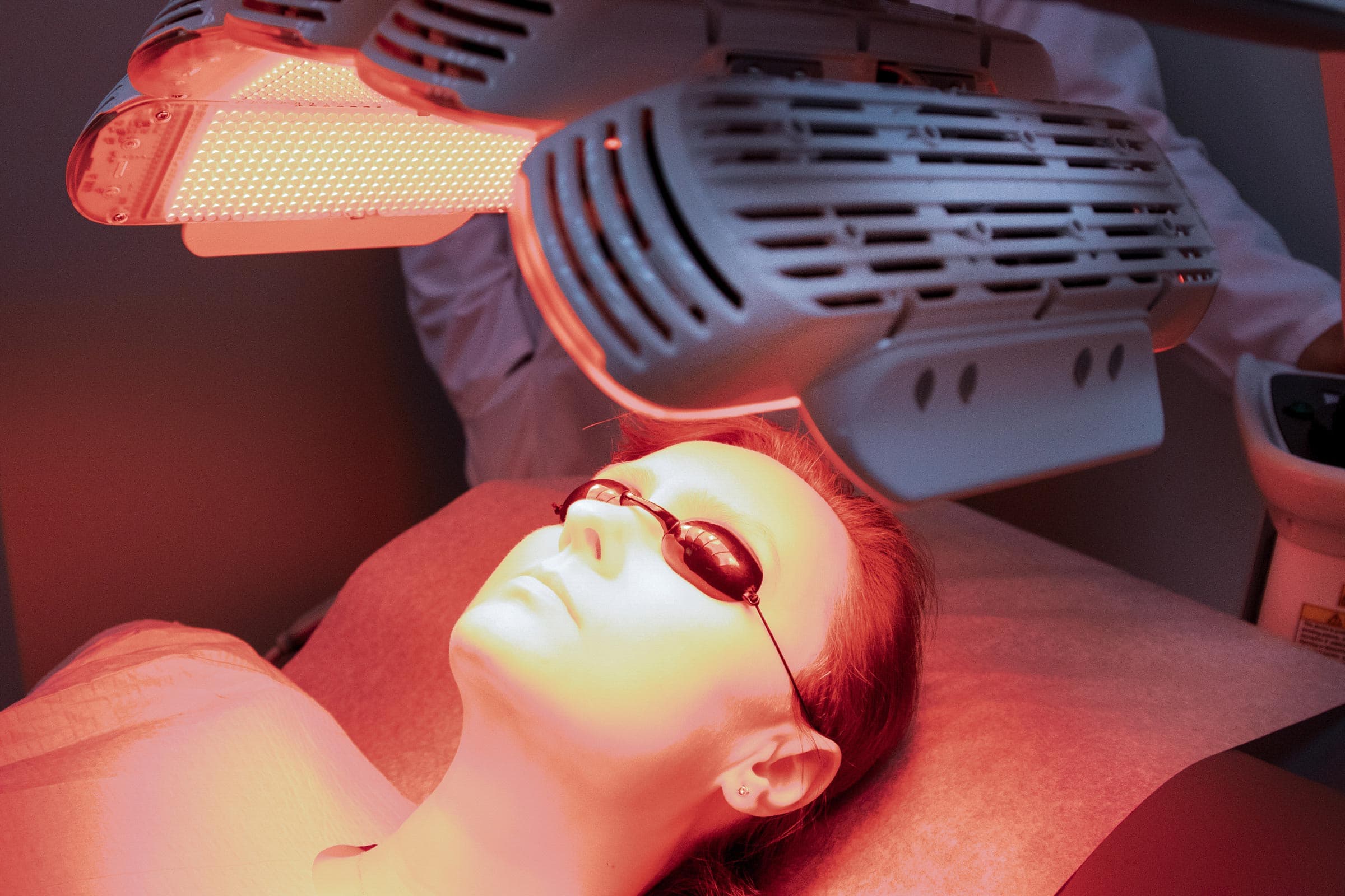
Intense pulsed light is not a laser. It emits non-coherent light across a broad band, typically between 500 and 1200 nanometres, which is then narrowed with cut-off filters. Because several chromophores are targeted at once, a single session can address both the brown pigment of lentigines and the background redness and telangiectasia that usually accompany photodamage. That breadth is its principal advantage over pigment-specific lasers.
In a randomised split-face trial comparing intense pulsed light with a Q-switched alexandrite laser for freckles and lentigines, the Q-switched laser achieved greater pigment clearance, while intense pulsed light produced fewer adverse pigmentary effects and less downtime [12]. The trade-off is characteristic of the field: greater selectivity generally brings greater efficacy and greater risk in the same instrument.
Intense pulsed light has also been studied in melasma. When added to a topical regimen in patients with refractory disease, it produced measurable improvement, but partial relapse was documented within six months and repeated treatment was required to maintain the result [13]. Intense pulsed light is generally avoided in Fitzpatrick skin types IV through VI, because the higher concentration of epidermal melanin competes for the delivered energy and raises the risk of burns, blistering, and post-inflammatory hyperpigmentation.
The Q-switched neodymium-doped yttrium aluminium garnet laser compresses its output into pulses measured in nanoseconds, which shatters pigment particles photomechanically rather than simply heating them. It operates at two wavelengths. The frequency-doubled 532 nanometre output is strongly absorbed by epidermal melanin and is well suited to solar lentigines and ephelides. The 1064 nanometre output penetrates more deeply and is absorbed less avidly by epidermal melanin, which makes it the safer wavelength in darker skin and the only practical option for dermal pigment. The same Q-switched Nd:YAG laser used for tattoo removal is the device applied to these pigmentary indications.
For discrete solar lentigines, the evidence is straightforward. A randomised controlled comparison of three lasers and liquid nitrogen found all four modalities effective, with the differences appearing in the rate of clearance and in the incidence of transient hypopigmentation and hyperpigmentation rather than in whether the lesions responded at all [14].
Melasma is where this laser is most often misapplied. Repeated low-fluence 1064 nanometre treatment, marketed as laser toning, does lighten melasma, and a randomised split-face trial of low-fluence Q-switched Nd:YAG against a low-fluence Q-switched alexandrite laser recorded improvement with both devices [15]. The problem is what follows. A case series documented mottled hypopigmentation and, in some patients, punctate leukoderma after repeated low-fluence sessions, a complication considerably harder to correct than the pigmentation being treated [16]. Laser therapy in melasma is therefore reserved for disease that has failed topical management, delivered conservatively, and always continued alongside photoprotection.
Picosecond devices shorten the pulse further, into trillionths of a second, which shifts the mechanism further toward photomechanical disruption and away from heat. A systematic review of picosecond lasers in dermatology found consistent evidence for benign pigmented lesions and tattoos, with the reduced thermal load offering a plausible advantage in patients prone to post-inflammatory hyperpigmentation, while noting that evidence in melasma remains limited and relapse remains common [17].
Chemical resurfacing removes pigment by removing the layers that contain it. A solution is applied under controlled conditions, the bonds between damaged keratinocytes are disrupted, and the epidermis is shed and replaced. Depth is governed by the agent, its concentration, the number of coats, and the contact time, and it is depth that determines both the result and the risk.
Superficial peels using glycolic acid, salicylic acid, or low-concentration trichloroacetic acid work at the level of the epidermis and are appropriate for lentigines, post-inflammatory hyperpigmentation, and epidermal melasma. In a comparative study in patients with darker skin, serial glycolic acid peels added to a topical regimen produced greater improvement in melasma than the topical regimen alone, although the difference did not reach statistical significance [18]. Medium-depth peels reach the papillary dermis and address more pronounced photodamage, at the cost of five to seven days of visible peeling and a higher risk of pigmentary complication.
The role of chemical resurfacing in pigmentation is adjunctive rather than primary. Peels are used to accelerate the response to topical therapy and to treat the diffuse dullness and textural change of photoaged skin that lasers targeting discrete lesions leave untouched. In melasma in particular, a peel performed without concurrent photoprotection and topical maintenance reliably fails.
Fitzpatrick skin types IV through VI require a different treatment logic, not simply lower settings. Epidermal melanin competes with the intended target for every photon delivered, which reduces efficacy and increases the risk of epidermal injury, and any epidermal injury in melanin-rich skin can itself produce prolonged post-inflammatory hyperpigmentation. Longer wavelengths, longer pulse durations, lower fluences, adequate epidermal cooling, and greater intervals between sessions are the standard adaptations [19].
Practical consequences follow. Intense pulsed light is generally unsuitable. The 1064 nanometre Nd:YAG wavelength is preferred over the 532 nanometre output. Test spots are performed before an area is treated. Topical regimens are used before and after the procedure to suppress the pigmentary response. Above all, expectations are set differently, because a slower and more conservative course is safer than a rapid one that provokes the exact problem being treated.
Solar lentigines and ephelides respond well and often clear substantially within one to three sessions, whichever modality is selected. They do not return in the same place if photoprotection is maintained, but new lesions continue to form on skin that continues to accumulate ultraviolet exposure. Treatment addresses the lesions present today, while sunscreen determines how many appear next year.
Melasma is managed rather than cured. Relapse rates are high, maintenance topical therapy and rigorous photoprotection are expected indefinitely, and devices are second-line tools that carry a genuine risk of worsening the condition when used aggressively. A treatment plan that promises permanent clearance of melasma in a fixed number of sessions is not describing the condition accurately.
Post-inflammatory hyperpigmentation improves as the underlying inflammation is controlled. Treating the acne or the dermatitis takes precedence over treating the pigment it left behind, since active inflammation regenerates that pigment as fast as any procedure removes it.
Facial pigmentation is one of the few areas of dermatology in which the diagnostic step and the treatment step are routinely reversed, with lasers applied to lesions that were never examined properly. At the Centre for Medical and Surgical Dermatology, pigmented lesions are examined by Dr. Maksym Breslavets with dermoscopy before any treatment is considered, and the modality is then matched to the diagnosis, the depth of the pigment, and the skin phototype. To have facial pigmentation assessed, request a consultation.
This article is intended for educational purposes and does not replace professional medical advice. Please consult your dermatologist for personalized recommendations.
Your feedback helps us improve our news and clinical insights
From medical dermatology to surgical procedures, our clinic provides comprehensive care for all skin, hair, and nail conditions.