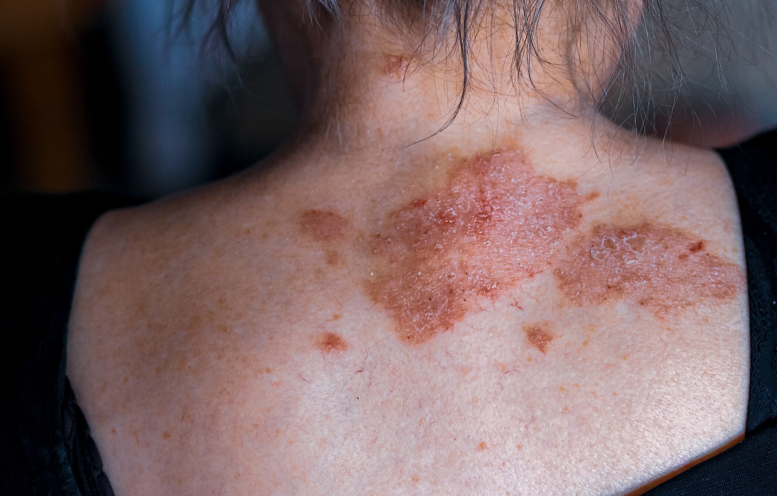
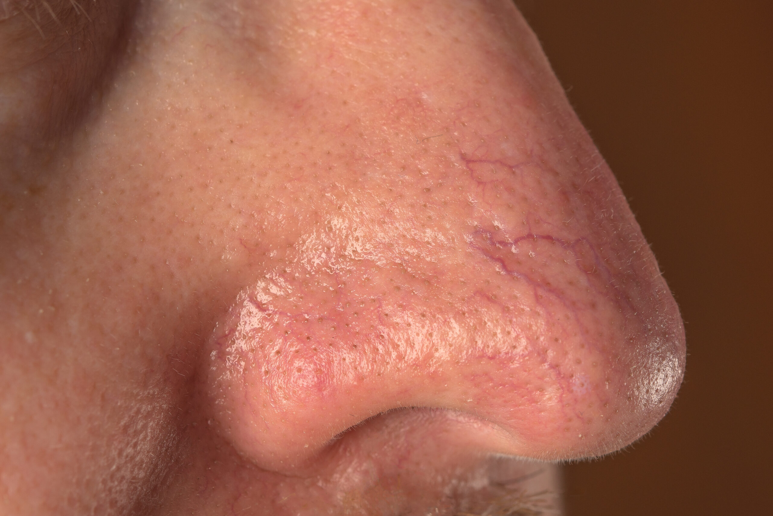
A melanocytic naevus, more commonly known as a mole, is one of the most frequently occurring benign skin lesion formed due to a local proliferation of melanocytes. Since a brown or black melanocytic naevus has a pigment melanin, it is often referred to as pigmented naevus.
A melanocytic naevus is generally classified as a congenital melanocytic naevus or an acquired naevus. A congenital melanocytic naevus is present at birth, while acquired naevus appear later in life.
Everyone has melanocytic naevus. Approximately 1% of the world’s population is born with at least one congenital melanocytic naevi. Individuals with fair skin have more melanocytic naevi in comparison with someone who has a darker skin type.
Moles that appear between 2 and 10 years of age are the most prominent and persistent throughout one’s life. Melanocytic naevi formed in later childhood and adulthood are usually caused due to sun exposure; they may eventually disappear.
The exact reason why local proliferation of naevus cell occurs is unknown. However, it has been determined that genetic factors, immune status, and sun exposure contribute to their formation. For example, individuals who have multiple melanocytic naevi have family members with similar skin lesions. Somatic mutations occurred in RAS genes are linked with congenital melanocytic naevi. New melanocytic naevi may arise due to taking BRAF inhibitor drugs like dabrafenib and vemurafenib.
Melanocytic naevi vary between different patients in terms of their clinical, dermatoscopic, and histological appearances. They can affect any part of the body. Their appearance depends on the body site they are found at. Melanocytic naevi can be flat or protruding, vary in colours (from pink or flesh tones to dark brown and black), and range in dimeter from a few millimetres to couple centimetres. Even though most of melanocytic naevi are have a round or oval shape; they sometimes may have irregular ones.
Melanocytic naevi is often associated with melanoma and if left untreated will result in death from skin cancer. Melanoma and a harmless melanocytic naevus may look alike. However, eventually melanoma changes its structure and increases in size. Patients who have a lot of naevi are in greater risk of developing melanoma. It is important to keep in mind that melanocytic naevi may enlarge, disappear, or regress for other reasons besides melanoma, such as pregnancy and sun exposure.
Melanocytic naevi are usually diagnosed clinically by their appearance. If abnormalities have been detected, further examinations will be done.
Majority of moles are harmless and left untreated. They may only be removed for cosmetic reasons, if a mole is a nuisance due to irritations caused by razor or clothing, or to exclude possibility of cancer.
Centre for Medical and Surgical Dermatology offers different melanocytic naevi treatments unique to each patient.


