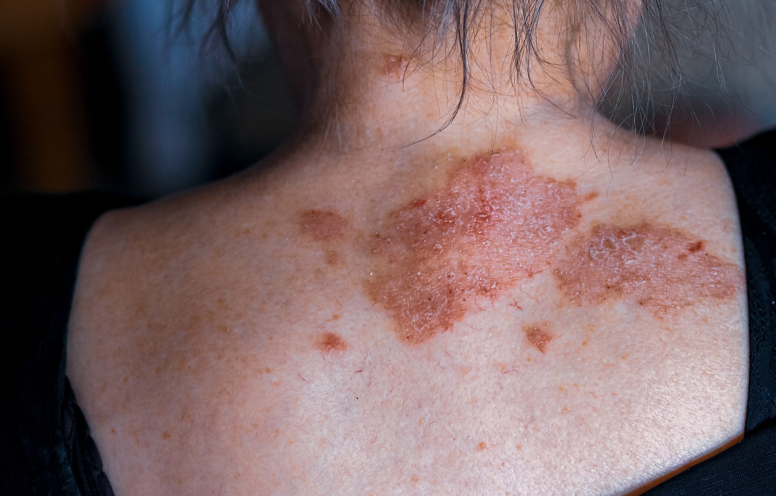
A melanocytic naevus, also known as a mole, is a very common benign skin lesion formed due to a local proliferation of melanocytes. Brown or black melanocytic naevus often called pigmented naevus because of melanin’s presence.
As mentioned in the previous post, melanocytic naevus is classified as a congenital melanocytic naevus or an acquitted naevus. By definition, a congenital melanocytic naevus is present at birth, while acquired naevus appears later in life.
Everyone has melanocytic naevus. Approximately 1% of the world’s population is born with at least one congenital melanocytic naevi. Individuals with fair skin have a higher prevalence of developing more melanocytic naevi in comparison with someone who has a darker skin type.
Moles that appear during ages 2 and 10 are the most prominent and persistent throughout an individual’s life. Melanocytic naevi formed in later childhood and adulthood are usually caused due to sun exposure and may disappear with time.
Melanocytic naevi are diagnosed clinically based on their appearance. The majority of moles are harmless and do not require any treatment. They may only be removed for cosmetic reasons or to exclude the possibility of skin cancer. Moles can also be removed if they are perceived as a nuisance due to getting irritated by a comb, razor, or clothing.
Moles are considered abnormal if they change in size, structure, shape, or colour. If a new naevus develops after the age of 40 years, it has to be assessed by dermatologist to exclude possibilities of melanoma. If a melanocytic naevus is bleeding, itchy, or crusty, additional examinations have to be done as well. Lastly, if ABCD (Asymmetry, Border irregularity, Colour variation, Diameter greater than 6 mm) characteristics are present, moles have to be assessed by a dermatologist for the presence of suspicious features.
Naevi that are considered suspicious for melanoma have to be excised for histopathology.
Surgical techniques used for removal of nevus include excision biopsy, shave biopsy, laser, or electrosurgery. Excision biopsy is performed on flat or suspicious melanocytic naevus. Shave biopsy is done on a protruding melanocytic naevus. Laser may be administrated to reduce pigmentation or remove coarse hair.
Development of melanocytic naevi can be minimized if conscious sun protection is maintained, especially if it is followed since birth. It helps to reduce skin ageing and development of skin cancer.
It is advised to choose fabrics that are designed for sun exposure (SPF 50+). Sunscreen has to be applied to uncovered areas. Other recommendations include wearing hair, long sleeves, a long skirt, or pants.
Majority of moles formed during childhood remain for the rest of one’s life. Teenagers and young adults have the biggest number of naevi present. With age, the number of naevi starts to decline because some moles fade away with time.
In order to increase the chances of detecting melanoma in the early stages, it is recommended to perform a monthly self-skin examination. If a patient is noticing changes in their mole(s) or formation of a new lesion, they should consult their dermatologist. It is highly recommended for patients with atypical looking naevi, many naevi, or past history of skin cancer to schedule regular skin check-ups with a dermatologist.
Centre for Medical and Surgical Dermatology offers different melanocytic naevi treatments unique to each patient.
For more information regarding the melanocytic naevi, visit the following link:
For more information regarding skin cancer and self-exams, visit the following link:
For more information regarding melanoma, visit the following link:
For more information regarding Dermato-oncology, visit the following link:


