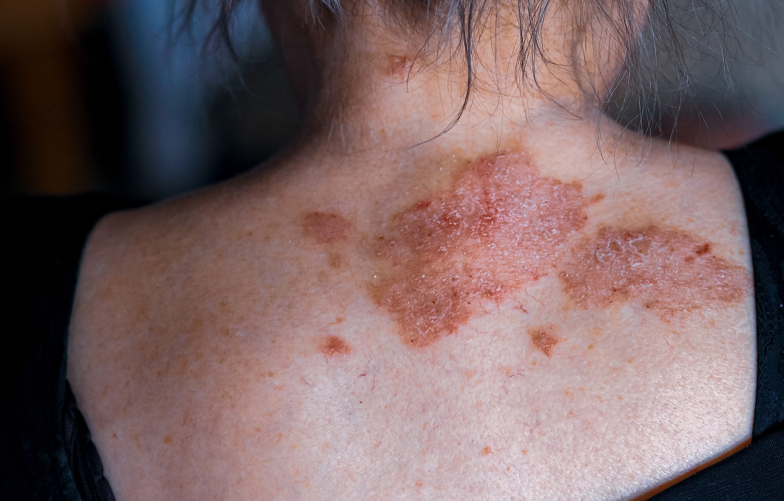
Excisional biopsy is the term used for describing the removal of skin lesion(s) by completely cutting them out of the affected site. The procedure is usually done under local anesthetic that is injected into the skin to make this area numb. The injection would sting transiently. After the procedure is done, a suture or dressing may be applied to the site where the biopsy was done.
There are different types of skin biopsies: punch biopsy, shave biopsy, curettage, incisional biopsy, and excision biopsy.
A punch biopsy is considered to be the most useful type of biopsy. It is quick to perform, convenient, and the procedure leaves a small wound. The sample of the skin has a full thickness which enables the pathologist to have a good overall overview of the epidermis, dermis, and subcutis.
A disposable skin biopsy punch is used to perform a punch biopsy. This tool has a round stainless steel blade ranging from 2-6 mm in diameter. The most common punches are 3 and 4 mm. The physician holds the instrument perpendicular to the skin site and rotates it in order to pierce the skin. With the help of forceps and scissors, the skin sample is subsequently removed.
Shave biopsy is used for superficial skin lesions in order to confirm the diagnosis of basal cell carcinoma, for example. A tangential shave of the skin is done with a scalpel (it is a special shave-biopsy instrument or razor blade). Stitches are not needed. The wound takes 1-3 weeks to heal.
The skin curette is used for scarping off the superficial skin lesion (e.g. seborrheic keratosis). It is important to note that the obtained samples are not used for determining if the lesion has been removed completely.
The incisional biopsy is used for removing larger and deeper parts of the skin with the help of a scalpel blade. Stitches are needed.
The excision biopsy is used for the complete removal of skin lesions like skin cancer. The margin of the surrounding skin is used for improving the chances of complete cancer removal. The smaller lesions are removed with a scalpel blade, while larger lesions are removed with a skin flap (moving adjacent skin on the wound site to cover it) or graft (skin is taken from another site to patch the wound).
Skin biopsy is usually a pretty straightforward process and chances of getting any post-procedure complications are uncommon. However, the larger the removed skin site is, the higher the chances are for possible complications.
For example, intraoperative and/or postoperative bleeding can occur in any patient. Bacterial wound infection occurs in about 1-5% of excision biopsies. It is very uncommon in small punch, shave, or incisional biopsies. The site of biopsy, ulcerated or crusted skin lesions, older age, diabetes, or usage of immunosuppressive medicines may increase the risk of possible wound infection.
The permanent scarring as a result of the post-biopsy is quite common. Some body sites, such as the center of the chest are prone to the development of excessive or hypertrophic scars.
After the biopsy was done, it is being sent to the pathology laboratory. It takes about 1 to 2 weeks to review the results.
Centre for Medical and Surgical Dermatology offers unique and personalized biopsy treatment options for each patient.
One Comment
Comments are closed.
Related Posts


Μy brother suggested I may like this blog. He was once totally right.
This pubⅼish truⅼy made my day. You cann’t
imagine juѕt how so much time I had sρent for this info!
Thanks!