Skin Cancer, Self-Exams, and Dermatoscopic Skin Surveillance
Discovering melanomas can be quite challenging with a naked eye for many patients. Throughout various skin cancer educational campaigns, some patients and their relatives gained more knowledge about possible skin cancer arising and became able to detect suspicious lesions. As a result, more cases have been reported to dermatologists, and treatment, if needed, began at early stages before the disease could progress further. However, in many cases, patients are unaware that some of their lesions are diagnosed as melanoma and become really surprised when dermatologists reveal the presence of skin cancer during a regular examination. Canadian Dermatology Association highly encourages individuals to report any changing and concerning lesions to their dermatologists for further assessment and clinical diagnosis. Moreover, any changes in asymptomatic (atypical) moles and the appearance of any new lesions on the skin in individuals 35 years old and older are considered as warning signs that cannot be ignored.
Studies have shown that an average person may have trouble accurately assessing one’s skin for the presence of any changes, especially if this change lasts for a prolonged period of time or if it is located at the site that cannot be easily seen (e.g. back).
In comparison with other more prevalent carcinomas, melanoma’s anatomical distribution is very unpredictable because it does not only appear on the areas of skin exposed to UV rays. However, due to popularity of UV tanning beds and full-body tanning sessions during summer, almost the entire body gets exposed to UV rays which increase the risk of developing melanoma skin cancer, especially in western societies.
Lastly, 71% of nevus-associated melanomas appear as new lesions on the skin and not in already existing moles. It is important to keep in mind that any new lesions are also very critical and in many cases, they go unnoticeable during self-skin exams.
Dermatoscopic Skin Surveillance refers to a special procedure of skin screening via dermatoscopy in order to assess moles and the presence of any high-risk malignant melanomas. The procedure is composed of a clinical full-body skin examination and assessment of concerning lesions. The purpose of the assessment is to detect any new and changing lesions.
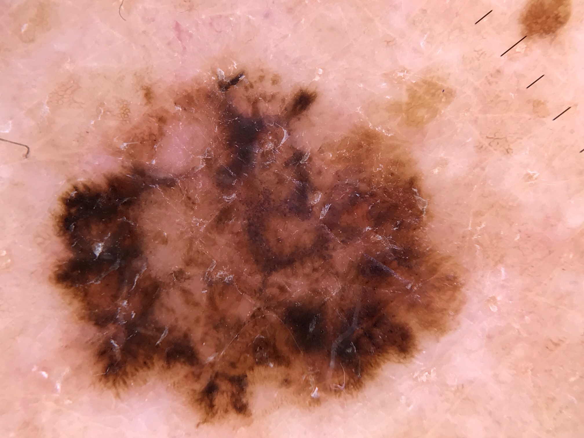
Dermatoscopy refers to skin examination via skin surface microscopy. The device contains a high-quality magnifying lens and a powerful lighting system. It enables the examination of different skin structures and patterns. The current market offers these hand-held devices with different lightweight and battery powers.
The following characteristics of skin lesions are assessed with the help of dermatoscope: symmetry or asymmetry, distribution of pigment (e.g. brown dots, lines, clods, and areas with undefined structures), homogeny/uniformity (sameness and repetition) or heterogeny (structural differences that appear on a particular lesion), skin surface keratin (small white fissures, crypts, and cysts), border of lesion (radical, sharply cut off, or fading streaks), presence of ulceration (full-thickness loss of epidermis), and vascular morphology (regular or irregular). As previously mentioned, dermatoscope is very essential for assessing various lesions like melanoma, atypical naevi, basal cell carcinoma, moles (benign melanocytic naevus), and seborrheic keratosis.
Dermatoscopic Skin Surveillance is useful for individuals who have many moles (>50-100), moles on the back, dysplastic or atypical naevi (large moles that appear in unusual shape and colour). Individuals who already have a history of melanoma and/or who have a history of melanoma running in their families are highly encouraged to perform regular skin exams. Lastly, individuals who have fair skin that experienced severe or repeated sunburns are advised to sign-up for a skin exam as well.
Dermatoscopic Skin Surveillance is extremely advantageous because melanoma and other pre-cancerous lesions can be detected at very early stages since new lesions and already existing ones can be detected with great accuracy. If dermatologist determines a concerning and pre-cancerous/cancerous lesion, it can be removed at earliest stage as possible which greatly decreases the risk of developing melanoma.
Centre for Medical and Surgical Dermatology treats any cancerous and pre-cancerous cases with extreme caution and a high degree of professionalism. Various treatment options for these cases are offered and are unique for each patient. For more information, visit the following links:
Non-melanoma skin cancer (basal cell carcinoma and squamous cell carcinoma)
Seborrhoeic keratosis (pre-cancerous condition)


