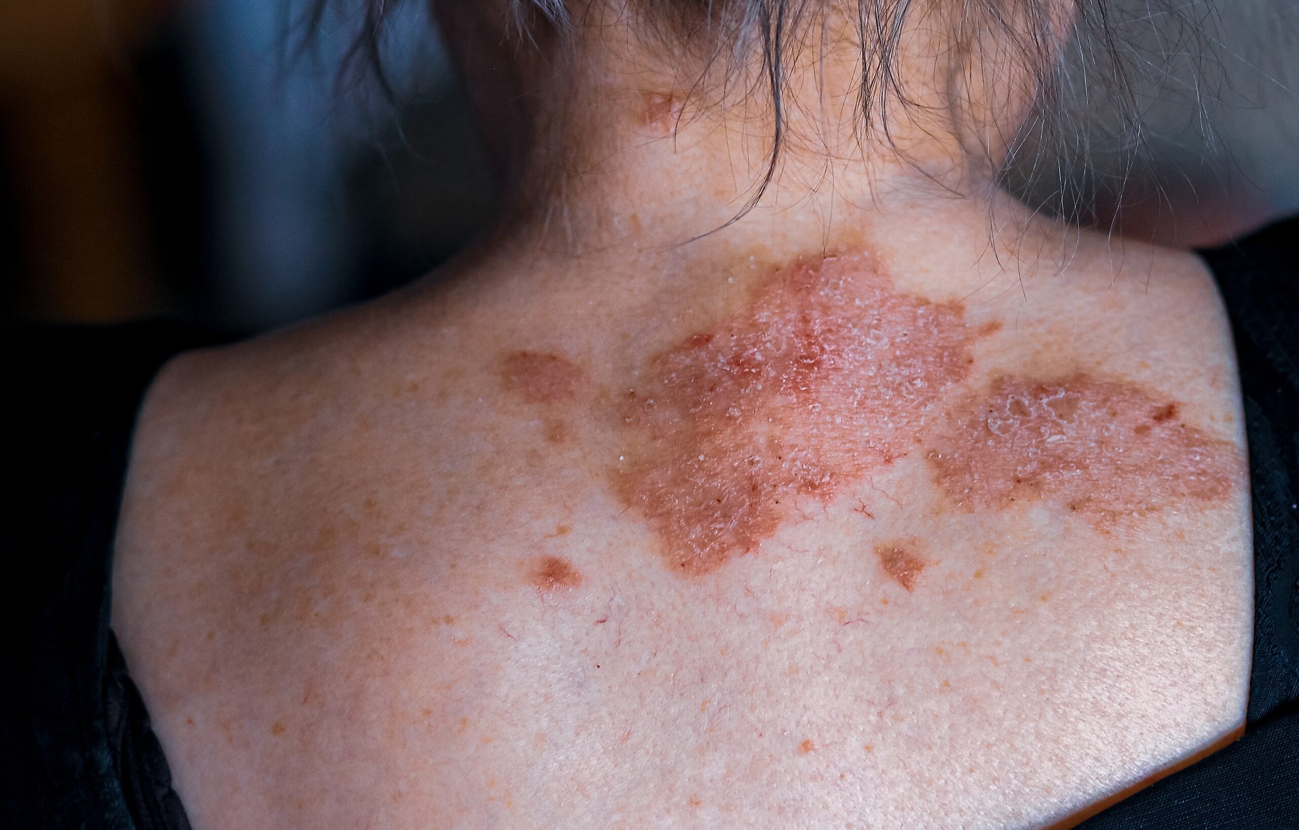
Seborrheic keratosis is a harmless, wart-like skin growth that commonly appears during adulthood as a sign of skin aging. Some individuals may have numerous seborrheic keratoses.
These growths, also known as SK, basal cell papilloma, senile wart, brown wart, wisdom wart, or barnacle, are part of a broader category of benign keratoses, which includes related scaly skin lesions like solar lentigo and lichen planus-like keratosis. Seborrheic keratoses are highly prevalent, with over 90% of adults over 60 having at least one. They can develop in people of all races, usually emerging in their 30s or 40s, and are rare in those under 20.
The exact cause of seborrheic keratoses is unclear. While the name suggests a link to seborrheic distribution and sebaceous glands, they do not adhere to these patterns. They are considered degenerative, becoming more numerous over time. Some individuals may have a genetic predisposition to develop a large number of them. Factors like sunburn, skin friction, viral infections, and genetic mutations can contribute to their formation. They can also develop from solar lentigo. Seborrheic keratoses appear on various skin areas but not on palms, soles, or mucous membranes.
Seborrheic keratoses exhibit diverse characteristics, appearing as flat or raised papules or plaques, ranging from 1 mm to several centimeters in diameter. They can be skin-colored, yellow, grey, light brown, dark brown, black, or mixed colors, and have a smooth, waxy, or warty surface. These growths may occur singly or in groups, often appearing in specific areas like the scalp, under the breasts, over the spine, or in the groin. While seborrheic keratoses are not precancerous, they can be challenging to distinguish from skin cancer, and in rare cases, they may coexist with skin cancer or indicate underlying internal malignancies.
Eruptive or irritated seborrheic keratoses can result from adverse reactions to medications or dermatitis. They may present as inflamed, red, and crusted lesions, sometimes leading to eczematous dermatitis.
The diagnosis of seborrheic keratosis is often straightforward, characterized by the presence of a raised, well-defined, warty plaque. While these growths usually have a distinct appearance, there are instances where they may resemble skin cancers like basal cell carcinoma, squamous cell carcinoma, or melanoma. Dermatoscopy, a skin examination method, often reveals an irregular structure in seborrheic keratosis, which is also a feature seen in skin cancer. Specific dermatoscopic clues, such as multiple orange or brown clods (caused by keratin within skin surface crevices), white milia-like clods, and curved thick ridges and furrows forming a brain-like or cerebriform pattern, can help differentiate them.
If uncertainty persists regarding the diagnosis, a partial shave, punch biopsy or diagnostic excision may be recommended to provide a definitive answer. The dominant histopathological characteristics of seborrheic keratosis include features like melanoacanthoma (deeply pigmented), acanthotic, hyperkeratotic or papillomatous, adenoid or reticulated, clonal or nested, adamantanoid or mucinous, desmoplastic, and irritated.
In terms of treatment, individual seborrheic keratoses can be easily removed if desired. Reasons for removal might include their unsightly appearance, itchiness, or the tendency to catch on clothing. Several methods can be employed for removal, including cryotherapy (using liquid nitrogen) for thinner lesions (which may require repeated sessions), curettage or electrocautery, ablative laser surgery, shave biopsy (removing with a scalpel), and focal chemical peel using trichloroacetic acid. It is important to note that each method has its disadvantages, with one concern being the potential for treatment-induced loss of pigmentation, especially in individuals with darker skin. Removing multiple lesions in a single session can also be challenging.
As for prevention, there is currently no known way to prevent the development of seborrheic keratoses. These growths often persist over time, although there are instances where individual or multiple lesions may spontaneously regress or improve through the lichenoid keratosis mechanism. In cases associated with dermatitis, regression may occur after effectively controlling the underlying skin condition.
Centre for Medical and Surgical Dermatology offers unique and personalized seborrheic keratosis treatment options for each patient.


