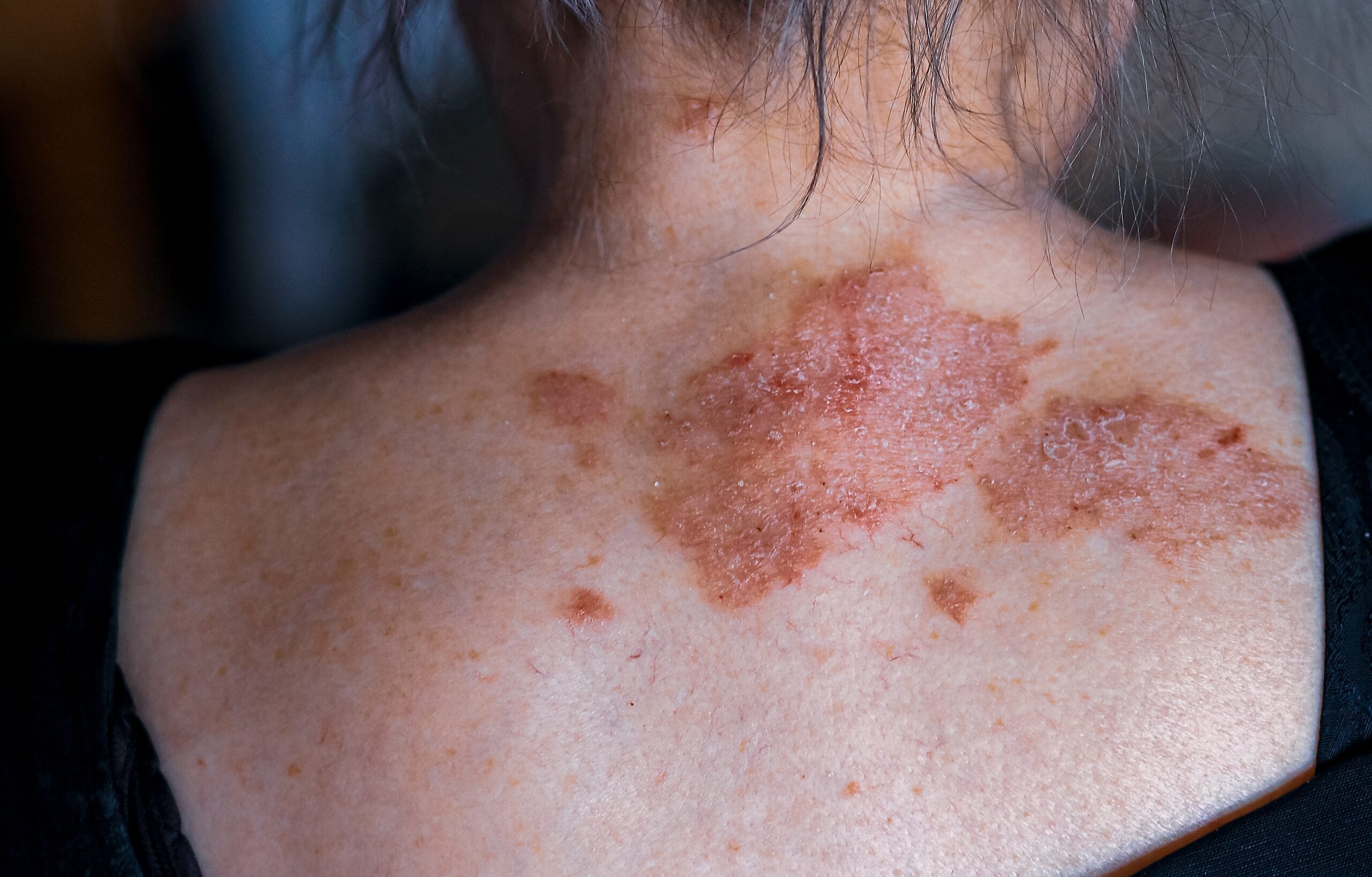
Nail psoriasis, also known as psoriatic nail dystrophy, occurs when psoriasis affects the nail matrix or nail bed, leading to specific and non-specific clinical changes in the nail. Psoriasis itself is a complex systemic disease involving inflammation and epidermal hyperproliferation.
Nail psoriasis is prevalent in individuals with chronic plaque psoriasis, affecting approximately 90% of them at some point in their lives. While it is more common in adults, with a prevalence of up to 80%, it is less frequently reported in children, occurring in 7–13% of cases. When psoriatic nail disease occurs independently without skin or joint involvement, it is observed in about 5–10% of adults.
Psoriatic nail disease is considered a potential risk factor for the development of psoriatic arthritis and is often found in association with severe cutaneous psoriasis that persists for a long time.
Nail psoriasis can affect anyone at any age, although there may be a higher incidence among males according to one significant case series.
The condition can involve various parts of the nail, such as the nail bed, nail matrix, hyponychium, and nail folds.
Several theories attempt to explain the causes of nail psoriasis, including the activation of the antimicrobial peptide LL-37 by Candida and the cytokine overflow theory, as well as increased expression of interleukin (IL)-10 in the affected nail bed compared to downregulation of IL-10 in psoriatic skin lesions. Additionally, psoriasis may be triggered by onychomycosis (fungal infection of the nail) or nail trauma.
Nail psoriasis may cause tenderness, pain, altered sense of fine touch, and difficulties with fine motor tasks like handling shoelaces or buttons.
Clinical signs of nail matrix involvement include pitting, leukonychia, red spots in the lunule, and onychorrhexis (longitudinal nail ridge, split, or fissure). Clinical signs of nail bed involvement include the oil-drop sign and salmon patch, onycholysis (with a pink zone proximally), subungual hyperkeratosis, and splinter haemorrhages under the distal third of the nail plate. Beau lines (transverse lines and ridges) and nail crumbling are other clinical signs of this condition.
Complications of nail psoriasis can include secondary onychomycosis in the damaged nail plate, psychosocial effects impacting social relationships and work-related activities, and associations with psoriatic arthritis and metabolic syndrome.
Diagnosing nail psoriasis is often based on clinical evaluation, particularly in patients with concurrent psoriatic arthritis and/or cutaneous psoriasis. The Nail Psoriasis Severity Index (NAPSI) can be used to estimate the severity of the condition by scoring clinical signs in each quadrant of the affected nails.
Fungal microscopy and culture of nail clippings should be performed to rule out onychomycosis, which may precede or complicate psoriatic nail dystrophy.
In some cases, a proximal nail matrix biopsy might be necessary to confirm the diagnosis, especially when signs of psoriasis are absent elsewhere or when only one nail is affected and other potential causes must be excluded. However, biopsy carries the risk of causing permanent nail deformities.
Centre for Medical and Surgical Dermatology offers various treatment options for psoriasis which are unique for every patient.
For more information about psoriasis in general, visit the following link:


