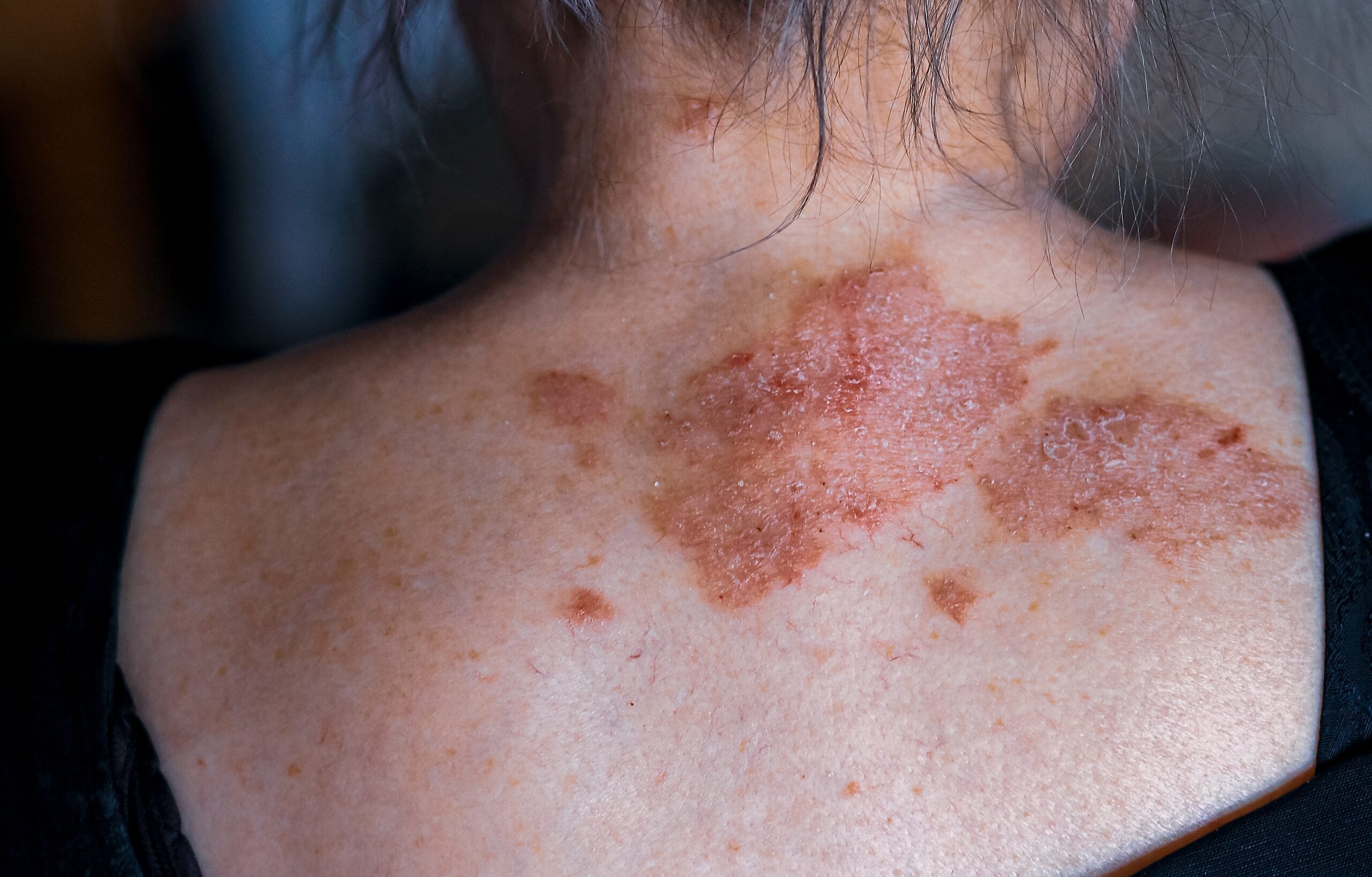
Melasma is a common acquired skin disorder characterized by bilateral, blotchy, brownish facial pigmentation. Previously known as chloasma—derived from the Greek word for “green”—the term melasma, meaning “brown skin,” is now preferred. Historically, it has also been referred to as the “mask of pregnancy.”
Melasma is more prevalent in women than in men, typically appearing between the ages of 20 and 40. It is most common in individuals who tan easily or have naturally brown skin (Fitzpatrick skin phototypes III, IV), and less common in those with fair skin (Fitzpatrick types I, II) or black skin (Fitzpatrick types V, VI).
The etiology of melasma is complex, often linked to photoaging in genetically predisposed individuals. The condition results from the overproduction of melanin by melanocytes, which is either taken up by keratinocytes (epidermal melanosis) or deposited in the dermis (dermal melanosis, melanophages).
Several factors contribute to the development of melasma. About 60% of individuals with melasma report having affected family members, indicating a significant genetic component. Ultraviolet (UV) and visible light stimulate melanin production, exacerbating the condition. Hormonal factors also play a role; pregnancy and the use of estrogen/progesterone-containing contraceptives, intrauterine devices, implants, and hormone replacement therapy are implicated in approximately one-quarter of affected women, and thyroid disorders can be associated with melasma as well. Additionally, certain cancer therapies and perfumed soaps, toiletries, and cosmetics may trigger phototoxic reactions that lead to melasma. Ongoing research is examining the roles of stem cells, neural and vascular factors, and local hormonal influences in promoting melanocyte activation, further contributing to the disorder.
Melasma manifests as bilateral, asymptomatic, light-to-dark brown macules or patches with irregular borders. Distinct patterns include:
– Centrofacial:Affects the forehead, cheeks, nose, and upper lip (excluding the philtrum); accounts for 50-80% of cases.
– Malar: Involves the cheeks and nose.
– Mandibular: Appears on the jawline and chin.
– Erythrosis Pigmentosa Faciei: Characterized by reddened or inflamed areas.
– Extrafacial: Affects sun-exposed areas such as the forearms, upper arms, and shoulders.
Melasma can be classified into three types based on the level of melanin in the skin.
Epidermal melasma is characterized by well-defined borders and a dark brown color. Under a Wood lamp, it appears more apparent. Dermoscopy reveals scattered islands of a brown reticular network with dark fine granules. Treatment for epidermal melasma generally responds well.
In contrast, dermal melasma has ill-defined borders and a color that ranges from light brown to blue-grey. Under a Wood lamp, there is no accentuation. Dermoscopy shows a reticuloglobular pattern, telangiectasia, and arciform structures. Treatment for dermal melasma usually yields a poor response.
Mixed Melasma is the most common type, featuring a combination of blue-grey, light, and dark brown colors. Treatment usually shows partial improvement.
Melasma can significantly affect quality of life due to its visible nature. Diagnosis is typically clinical, supported by examination with a Wood lamp and dermatoscope. Occasionally, a skin biopsy may be required, revealing features such as melanin deposition in basal and suprabasal keratinocytes, intensely pigmented melanocytes, and increased blood vessels.
Conditions that may resemble melasma include:
– Post-inflammatory hyperpigmentation
– Solar lentigo and other forms of lentigines and freckles
– Acquired dermal macular hyperpigmentation
– Drug-induced hyperpigmentation
– Naevus of Ota and Naevus of Hori
Treating melasma often requires a combination of measures. General measures include year-round, lifelong sun protection with broad-brimmed hats, SPF50+ sunscreens containing iron oxides, and sun-smart behavior. It is also advisable to discontinue hormonal contraception if possible. Cosmetic camouflage can be used to cover the pigmentation.
Topical therapy is essential in treating melasma. The most effective formulation is a combination of hydroquinone, tretinoin, and moderate potency topical steroids, which can clear or improve 60-80% of cases. Other topical agents used alone or in combination include azelaic acid, kojic acid, cysteamine cream, ascorbic acid, methimazole, tranexamic acid, glutathione, and soybean extract.
Oral treatment options are also available. Tranexamic acid, which inhibits the conversion of plasminogen to plasmin, shows promise in treating melasma. Additionally, new oral treatments are currently being trialed.
Procedural techniques such as chemical peels and lasers can be used with caution, as they carry risks of worsening melasma or causing post-inflammatory hyperpigmentation. Superficial epidermal pigment can be removed using alpha-hydroxy acids (AHAs) like glycolic acid or beta-hydroxy acids (BHAs) like salicylic acid. Microneedling, intense pulsed light (IPL), and various lasers require expert application due to high risks of relapse.
Melasma can be challenging to treat and often responds slowly, particularly if it has been present for a long time. Even successful treatment may be followed by recurrence upon sun exposure. The chronic nature and risk of relapse necessitate lifelong sun protection and setting realistic treatment goals.
Overall, melasma requires a multifaceted treatment approach and ongoing management to mitigate its effects and improve patient quality of life.
Centre for Medical and Surgical Dermatology offers various treatment options for melasma which are unique for every patient. For more information on melasma condition, visit the following link:


