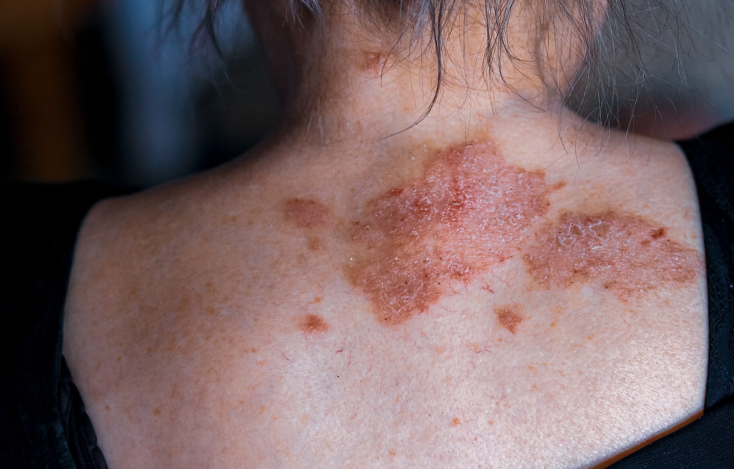
Naevi, more commonly known as moles, are referred to as pigmented and non-pigmented benign growth of the skin containing nevus cells and are very prevalent among the majority of adults. They come in different shapes: smooth, flat, elevated, pedunculated, hair or non-hair bearing. It is a common procedure done by dermatologists to remove the naevi for health or for aesthetic purposes.
To begin with, each lesion needs to be identified as ominous or malignant presentation based on ABCDE scale. It is an acronym that stands for:
- Asymmetry
- Border irregularity
- Colour variation
- Diameter greater than 6 mm
- Evolving or changing
After dermatologist has done a skin assessment and detected any changes with respect to ABCDE rules, lesion that are perceived as evolving and; thus, suspicious, would require a diagnostic biopsy.
The mole removal for the aesthetic purposes can be done in several ways.
One of the most promising techniques is radiofrequency surgery or radiosurgery.
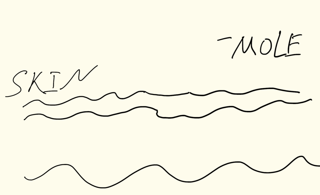
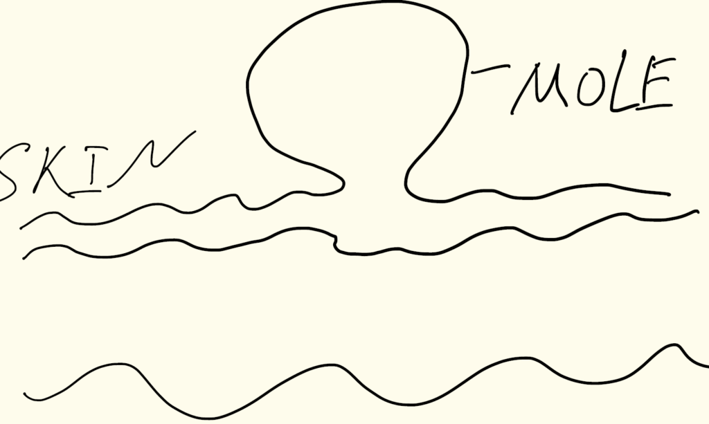
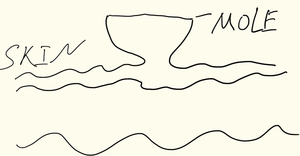
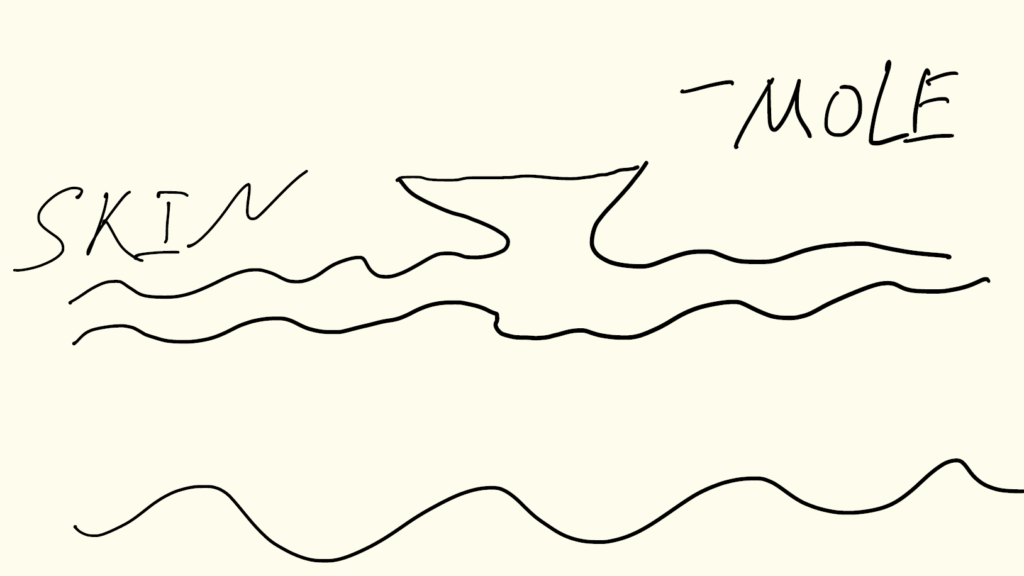
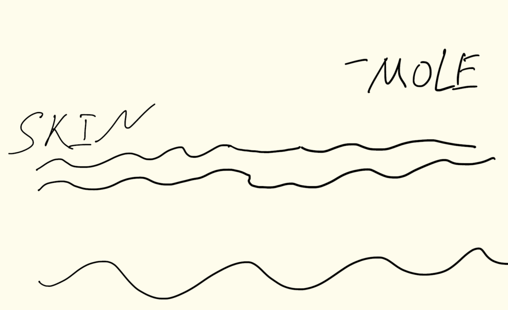
The method of the radiosurgery is based on the physical phenomenon of dissecting (cutting) the soft tissue like a skin with an energy of the radiowaves that are delivered to the tissue via a special electrode.
Prior to the removal, all lesions are anaesthetized with a local anesthetic, such lidocaine that makes the surgical procedure virtually painless.
The technique of lesion removal is described as “light and pressureless” ; the tip of the electrode is moving with barely any resistance resulting in a “melting” of the nevus. It is very precise and allows to remove even layers of skin in steps less than 1/10 of mm.
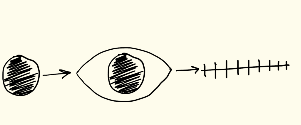
The target tissue is removed gently in multiple steps.
The steps are repeated until the lesion gets completely removed.
After radiofrequency surgery or radiosurgery procedure, it is highly recommended to apply medical grade petrolatum or special silicone based dressing gel on the lesion for about a week. It will allow the affected skin to gain its epithelized properties of being smooth, pink, and dry compared to the original wound. The lesion will be pink with some gradual color change back to normal which takes several weeks on average. In patients with extremely sensitive and fair skin tone, the lesion may remain pink for a couple of months. After the mole removal, a patient is scheduled for an 8-week follow-up appointment for the post-surgery check-up.
Another way to remove a mole is shave excision with a flexible blade.
The third option is to excise the lesion with a scalpel and close it with a stitches.
Centre for Medical and Surgical Dermatology offers mole removal procedures in different ways unique to each patient. For more information, visit the following link:
For more information regarding melanocytic naevi, visit the following links:
3 Comments
Comments are closed.
Related Posts

Very informative and detailed post!
I highly recommend reading it before considering mole removal.
Bravo, magnificent idea
Great admin work!