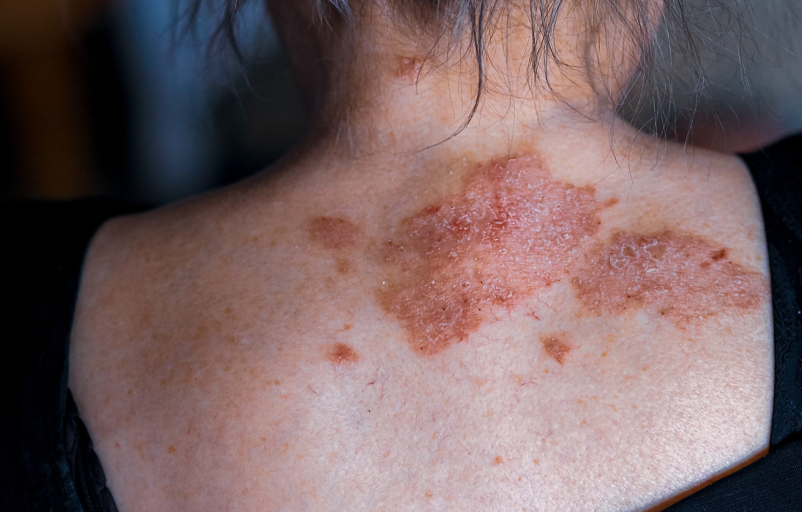
Erythroderma, a severe dermatological condition, is characterized by widespread, intense skin reddening due to inflammatory diseases. This condition often leads to exfoliation, or skin peeling, and is sometimes referred to as exfoliative dermatitis (ED). Affecting individuals of all ages and races, erythroderma is more prevalent in males, occurring three times as frequently as in females. Its emergence is typically linked to pre-existing skin disorders or systemic diseases, with around 30% of cases being idiopathic, meaning their cause is unknown.
In terms of demographics, erythrodermic atopic dermatitis primarily impacts children and young adults, while other forms of erythroderma are more common among the middle-aged and elderly population. The most frequent causes of erythroderma include drug reactions, various types of dermatitis (notably atopic dermatitis), psoriasis (particularly following the cessation of certain treatments), and pityriasis rubra pilaris. Other, less common causes encompass different dermatitis forms such as contact and stasis dermatitis, blistering diseases like pemphigus, cutaneous T-cell lymphoma (Sezary syndrome), and rare congenital conditions.
Moreover, erythroderma can be a manifestation of systemic illnesses, including haematological malignancies (e.g., lymphoma and leukaemia), internal cancers (e.g., rectal and lung carcinoma), graft-versus-host disease, and HIV infection. The progression of some skin diseases to erythroderma remains unclear, but it involves complex interactions between keratinocytes, lymphocytes, adhesion molecules, and cytokines, leading to rapid epidermal cell turnover.
Clinically, erythroderma often starts with a measles-like eruption, dermatitis, or plaque psoriasis, and can develop rapidly in acute cases or gradually in chronic instances. The condition is defined by widespread erythema and oedema, affecting over 90% of the skin surface. Patients experience significant discomfort, including intense itching, and can develop symptoms like swollen eyelids (ectropion), skin scaling, and changes in the nails and lymph nodes. The underlying cause might be indicated by specific clinical signs, such as serous ooze in atopic erythroderma or the persistence of psoriatic plaques.
Systemic symptoms can arise from either the erythroderma itself or its underlying cause, potentially leading to lymphadenopathy, hepatosplenomegaly, abnormal liver function, and fever, hinting at drug hypersensitivity or malignancy. Erythroderma’s complications are both acute and chronic, with patients experiencing temperature dysregulation, fluid and electrolyte imbalances, high-output heart failure, secondary skin infections, and potential systemic effects like pneumonia and hypoalbuminemia.
Diagnostic evaluations include blood tests revealing anomalies like eosinophilia, which may suggest lymphoma, and other tests to assess albumin levels and liver function. Skin biopsies are performed for unclear causes, showing nonspecific inflammation but can have diagnostic features in certain cases. Direct immunofluorescence is helpful if autoimmune blistering or connective tissue diseases are suspected.
Erythroderma represents a condition that carries significant potential for severity, even posing a life-threatening risk. In such cases, hospitalization is often necessary to facilitate continuous monitoring and address critical aspects, such as fluid and electrolyte balance, circulatory status, and body temperature.
Several general measures are recommended in managing erythroderma. It is essential to discontinue all unnecessary medications to prevent potential exacerbation of the condition. Simultaneously, close monitoring of fluid balance and body temperature is crucial for the overall well-being of the patient.
Moreover, maintaining skin moisture is a key aspect of erythroderma management. This involves the application of wet wraps, various wet dressings, emollients, and mild topical steroids to ensure optimal skin care.
Due to the heightened susceptibility to bacterial infections associated with erythroderma, prescribing antibiotics is a fundamental component of the therapeutic approach.
Additionally, the use of antihistamines, though variable in efficacy, is considered a potential strategy to alleviate the accompanying itch.
In instances where a specific cause for erythroderma can be identified, targeted treatments are recommended. For example, in cases of atopic dermatitis, initiating topical and systemic steroids may be warranted. Similarly, for psoriasis, treatment with acitretin or methotrexate is advised. These tailored interventions aim to address the underlying cause and enhance the overall effectiveness of the therapeutic regimen.
In most instances, the occurrence of erythroderma is difficult to prevent. Individuals with a documented drug allergy should be advised to abstain from the implicated drug permanently, and those who experienced a severe reaction are recommended to wear a drug alert bracelet. Updating medical records becomes crucial in the event of an adverse reaction to a medication, serving as a reference when initiating new drug therapies.
Patients grappling with severe skin conditions should be made aware of their potential susceptibility to erythroderma. They should receive education about the risks associated with discontinuing their prescribed medications and the likelihood of recurrence.
Regarding the prognosis of erythroderma, it hinges on the underlying disease process. If the causative factor can be eliminated or rectified, the prognosis is generally favourable. When erythroderma stems from the widespread dissemination of a primary skin disorder like psoriasis or dermatitis, it typically resolves with appropriate treatment of the skin ailment but may reappear unpredictably.
The course of idiopathic erythroderma is marked by uncertainty, with the condition possibly persisting for an extended duration and experiencing periods of acute exacerbation.


