Epidermolysis bullosa (EB) with congenital absence of skin, once known as Bart syndrome and also referred to as ‘type VI aplasia cutis congenita and epidermolysis bullosa’, was first identified by Bruce Bart in 1966. He observed this condition in a large six-generation family, characterized by symmetrical congenital skin absence on the lower legs, acral skin blistering, sometimes extending to mucous membranes and nail dystrophy. In 1986, Ilona Frieden introduced a classification for aplasia cutis congenita, organizing conditions into nine categories based on lesion location and the presence of other anomalies. This system placed Bart syndrome as type VI, highlighting its distinct combination of localized congenital skin absence, epidermolysis bullosa, and nail dystrophy.
A 2014 expert panel on EB proposed reclassifying the condition based on molecular characteristics, advocating for descriptive terminology over eponymous names, thus recommending the term EB with congenital absence of skin. The prevalence of this condition is rare, with aplasia cutis congenita occurring in 1–2 per 10,000 births, though the exact incidence of EB within this classification remains unclear.
EB with congenital absence of skin is noted for its autosomal dominant inheritance pattern, though sporadic cases have been documented. Its aetiology is multifaceted, affecting various skin membranes, which can be epidermal (simplex), junctional, or dermal (dystrophic). The initial family studied by Bart exhibited dystrophic EB, with ultrastructural changes in the dermal anchoring fibrils linked to the COL7A1 gene on chromosome 3, which encodes collagen type VII.
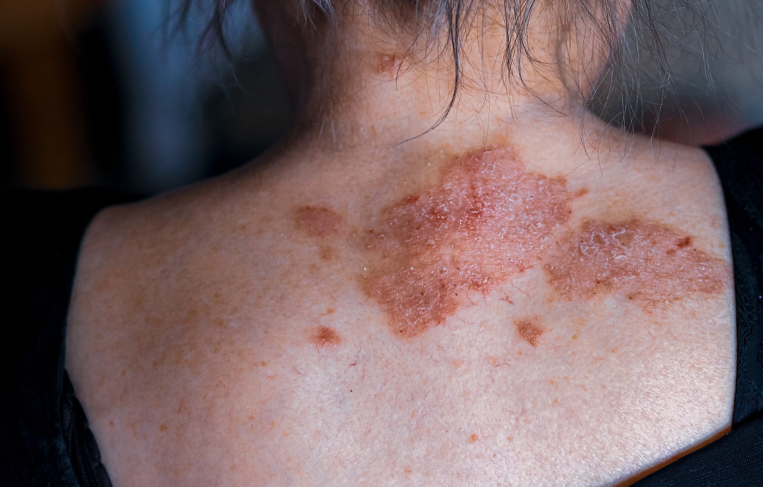
This condition is believed to result from in-utero trauma and skin fragility, leading to a symmetrical skin absence on the lower legs, which may rub together. EB with congenital absence of skin manifests as a clinical triad: congenital skin absence on the lower legs, any EB type elsewhere on the body, and nail changes ranging from absence to dystrophy. Typically, the absent skin areas are symmetrical and bilateral, and appear as sharply defined, glistening red ulcers.
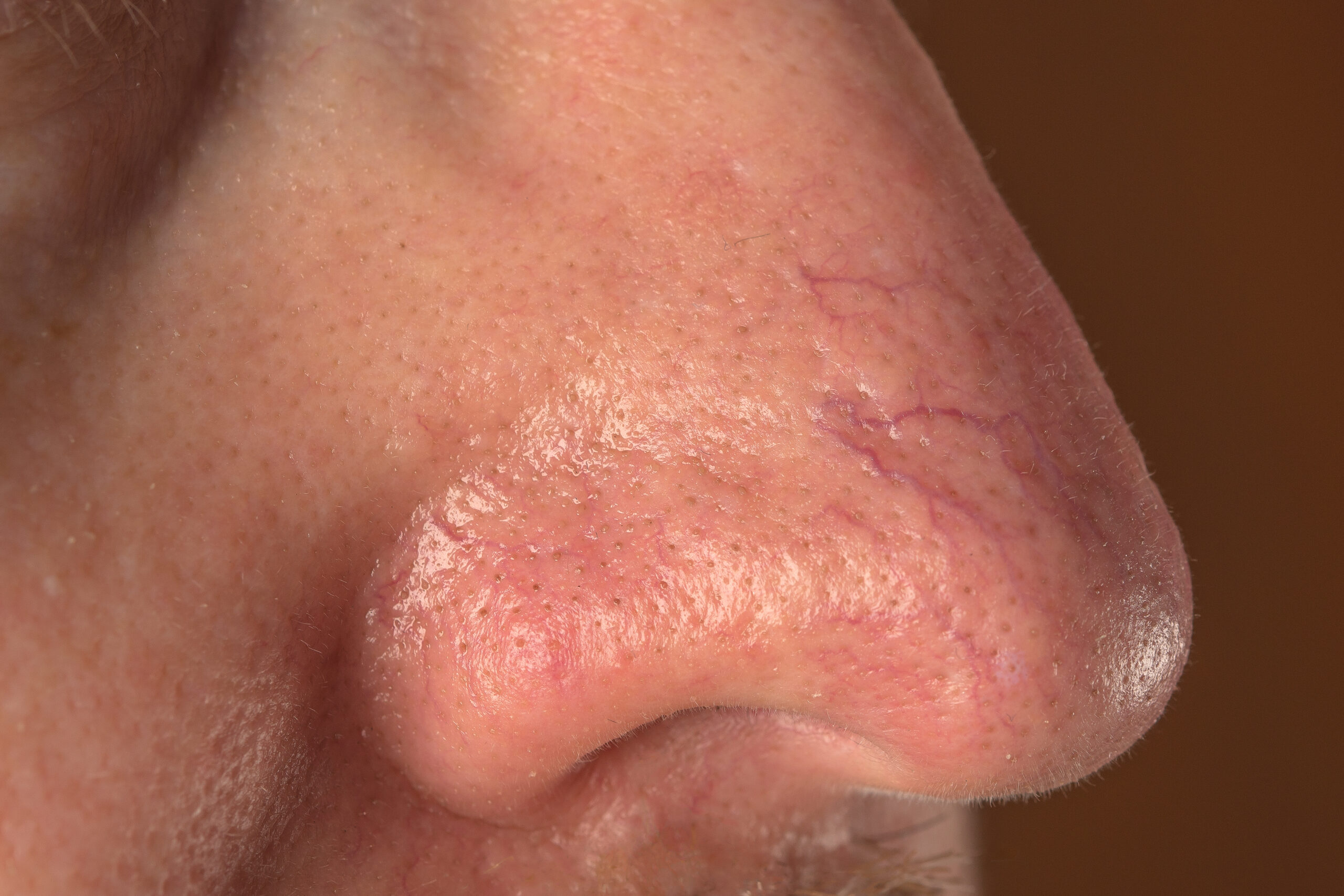
The original family’s phenotype varied, not all members exhibited the complete triad. Associated anomalies can include pyloric atresia, underdeveloped ears, a flattened nose, a broad nasal root, and wide-set eyes, with potential complications like infection, haemorrhage, hypothermia, hypoglycemia, and fluid balance disorders. Severe cases, particularly those with extensive mucosal involvement, have led to early fatalities.
Diagnosis is clinical, confirmed by histological skin examination to determine the EB type. Some cases show a subepidermal blister with a dermal inflammatory infiltrate, while electron microscopy in Bart’s original cases revealed a split below the lamina densa, indicative of dermal dystrophic EB. Other findings include dermal–epidermal junction separation and basal lamina interruption, similar to junctional EB, or splits above the basement membrane for epidermal simplex EB.
Differential diagnoses encompass other aplasia cutis congenita forms, other EB types, and Adams–Oliver syndrome, characterized by scalp aplasia cutis congenita, limb transverse defects, and skin mottling. Management primarily involves conservative treatment, focusing on healing, infection prevention, and scar minimization, with topical antibacterial ointments and wet dressings as standard care. Surgical intervention may be necessary for extensive defects.
The prognosis generally appears positive, with most congenital skin absence areas healing within weeks to months without long-term issues. However, follow-up with Bart’s original family revealed ongoing blister formation, milia, and mild scarring at blister sites, with atrophic hairless scars where the skin had been absent and no nail recovery.


