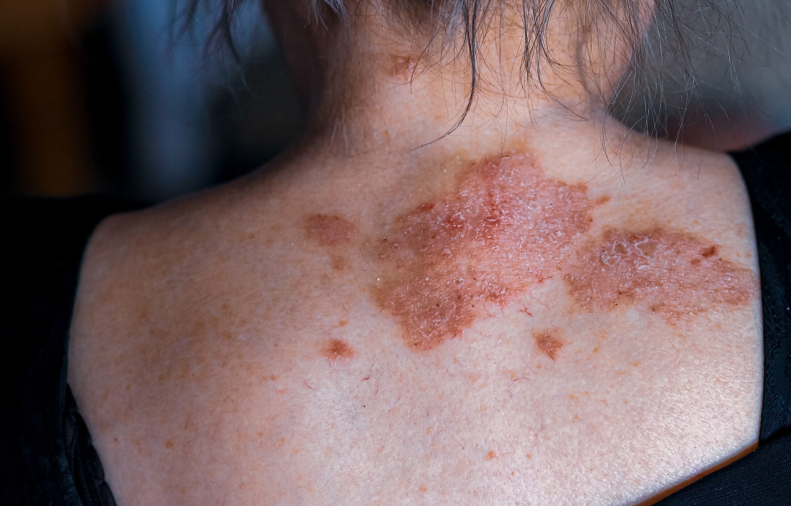
Eczema is a prevalent skin condition with several distinct clinical presentations, distinguished histologically by a spongiotic tissue reaction pattern. The terms eczema and dermatitis are frequently used interchangeably to indicate a diverse inflammatory reaction pattern affecting both the epidermis and dermis. However, ‘dermatitis’ denotes skin inflammation and is not synonymous with eczematous processes. A consensus among dermatopathologists suggests replacing ‘eczema’ with ‘spongiotic dermatitis’ to accurately describe the underlying histopathologic changes in so-called ‘eczemas’.
The spongiotic tissue reaction pattern involves intercellular swelling within the epidermis (spongiosis). Initially, there is an enlargement of spaces between keratinocytes and elongation of intercellular connections. Increased fluid accumulation leads to intraepidermal vesicle formation. Spongiotic dermatitis is a dynamic pathological process where vesicles may appear and disappear, situated at various epidermal levels. Epidermal infiltration by lymphocytes (exocytosis) is common. Parakeratosis develops above spongiotic areas, potentially due to the faster movement of keratinocytes toward the surface. Plasma droplets accumulate in parakeratosis mounds. Dermal alterations encompass varying degrees of oedema and a superficial perivascular infiltrate containing lymphocytes, histiocytes, and occasional neutrophils and eosinophils.
Clinically, eczema is categorized by aetiology. Histologically, eczema is better classified by chronicity. There are three histological stages of eczema: acute, subacute, and chronic. Eczematous conditions can start at any stage and transition to another.
Acute spongiosis is marked by extensive intercellular epidermal oedema, with widened spaces, desmosome disruption, and microvesicle formation. Vesicles are usually intraepidermal, but extensive vesiculation may lead to subepidermal vesicles. These vesicles contain proteinaceous fluid with lymphocytes and histiocytes. Allergic/contact dermatitis might involve eosinophils (eosinophilic spongiosis).
Subacute eczema is the most common form of spongiotic dermatitis. It presents mild to moderate spongiosis and exocytosis of inflammatory cells. Irregular acanthosis and parakeratosis are additional features. Superficial dermal perivascular lymphohistiocytic inflammation, endothelial cell swelling, and papillary dermal oedema are observed.
In chronic spongiotic dermatitis, mild spongiosis might be challenging to discern. Vesiculation is rare. Significant epidermal acanthosis often displays a psoriasiform pattern with hyperkeratosis, hypergranulosis, and limited parakeratosis. Papillary dermal fibrosis may be present.
There are multiple varieties of eczema, which will be discussed below.
Atopic dermatitis is common, particularly in children, associated with personal/family atopy history. Chronic spongiosis is more prevalent than acute or subacute patterns. A follicular pattern, showing spongiosis of follicular infundibula and sparse dermal infiltrate (follicular spongiosis), is notable.
Irritant contact dermatitis is provoked by water, detergents, and chemicals, irritant contact dermatitis commonly occurs on hands. Mild spongiosis, epidermal cell necrosis, and neutrophilic epidermal infiltration are typical histological features.
Sensitization to tolerated contacts triggers allergic contact dermatitis. Subacute, chronic, or acute patterns can emerge. The dermal infiltrate mainly contains lymphocytes and other mononuclear cells.
Chronic eczema, discoid or nummular dermatitis, is marked by papules evolving into coin-shaped patches. Features vary with chronicity.
Impaired venous circulation causes stasis dermatitis, common on legs. Mild spongiosis, parakeratosis, and scale crust are typical, with neovascularization, haemosiderin deposition, and fibrosis.
Precise diagnosis relies on clinicopathologic correlations, and pathologists should be specific while clinicians provide vital clinical data.
When conducting a skin biopsy for hand dermatitis, it is important to use special stains to check for fungal elements, aiming to rule out a potential dermatophyte infection (tinea). The histopathological characteristics that define spongiosis are also present in various other skin conditions that are not traditionally categorized as ‘eczema’. This complexity contributes to the challenge of defining the condition. This includes examples like pityriasis rosea, Gianotti Crosti syndrome, annular erythemas, miliaria, Grover disease, polymorphous light eruptions, papular urticaria, lichen striatus, and some pigmented purpuras. Determining the exact type of spongiotic dermatitis relies on a precise correlation between clinical and pathological information. For an accurate evaluation of the biopsy, the pathologist must be aware of the lesion’s location, distribution, patient’s age, and clinical morphology. Factors like lesion type (papules, vesicles, bullae, plaques, or patches), itchiness, presence of excoriations, duration of lesions, and response to standard treatment are significant.
In many cases, pathologists can only diagnose a general category of ‘spongiotic dermatitis, not otherwise specified’. They should be cautious about diagnosing an eruption as spongiotic or eczema if the pathological changes actually represent a different spongiotic condition. For instance, if lesions aren’t itchy, other diagnoses should be considered. If capillaritis is observed, it might indicate a pigmented purpura, warranting an iron stain. To exclude a dermatophyte infection, a special stain for fungal forms (like PAS or Grocott) is advisable in most cases. When eruptions are located on extremities in younger individuals, Gianotti Crosti syndrome is a possibility. If lesions are widespread and cause itchiness with papulovesicles, the biopsy should be examined in sections to identify conditions like Grover disease (which displays acantholysis) or papular urticaria (characterized by numerous eosinophils between cells).
Frequently, histopathological examination does not provide a definitive explanation of the cause or origin. Pathologists should aim for specificity while referring physicians play a vital role in offering relevant clinical details.
Centre for Medical and Surgical Dermatology offers various treatment options for eczema which are unique for every patient. For more information on this condition, visit the following link:


