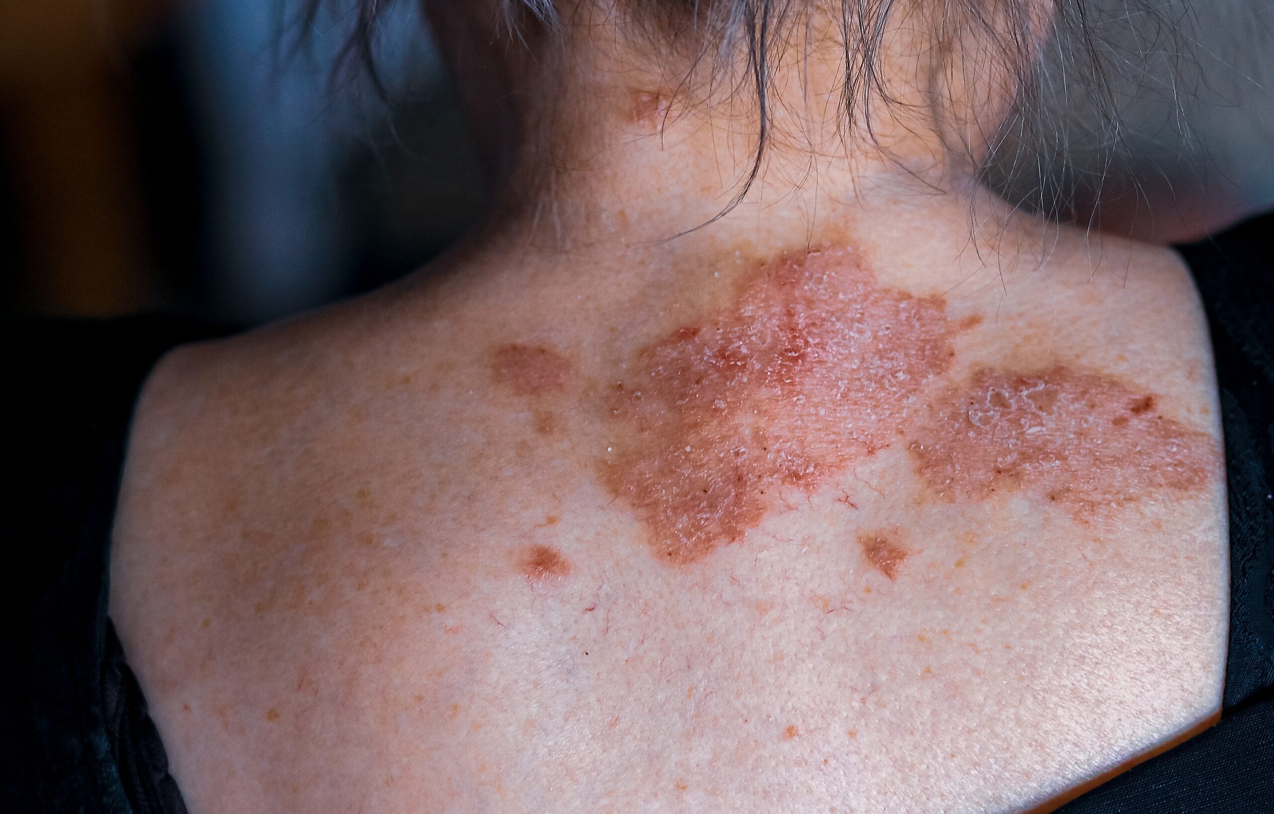
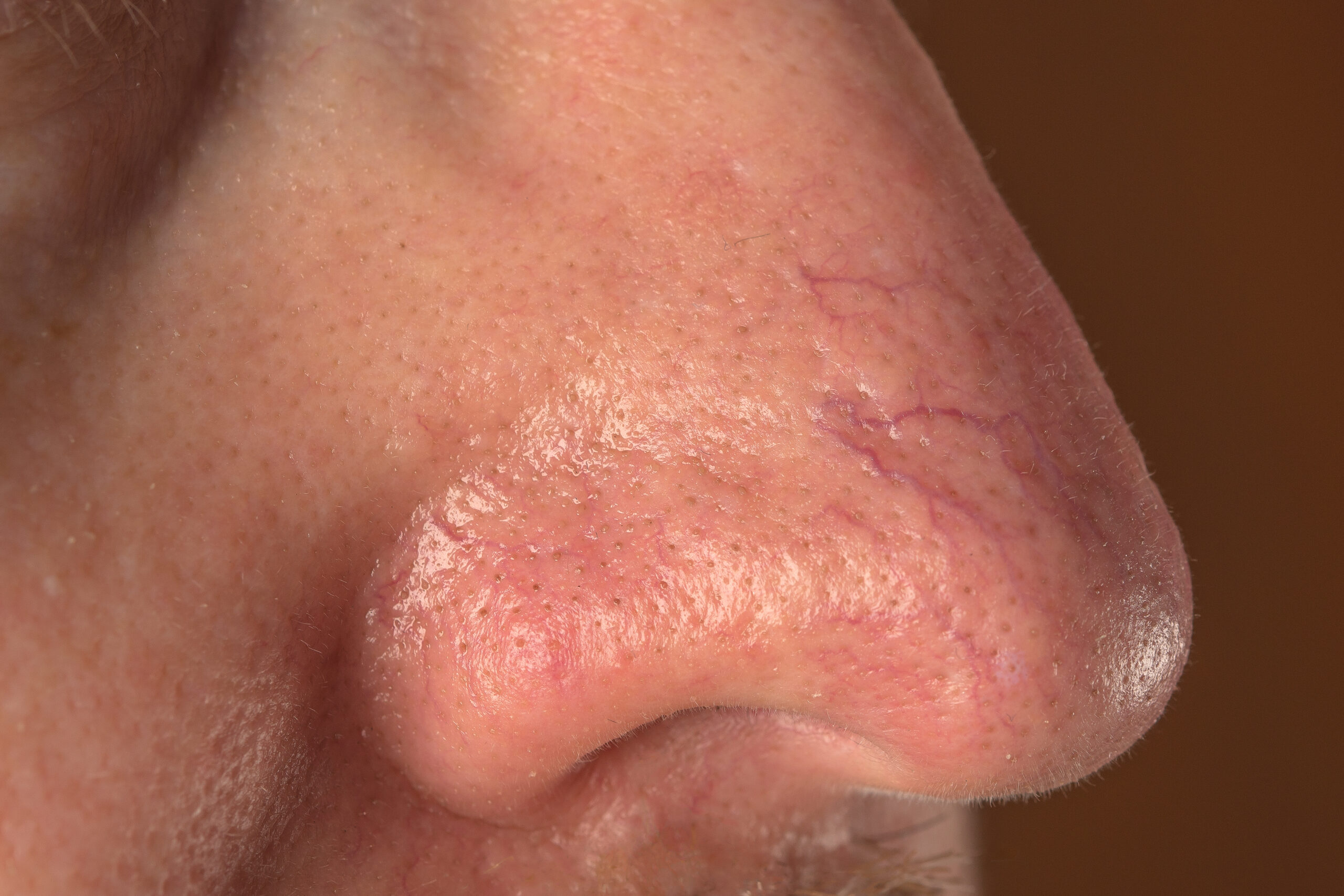
Cherry angiomas, also referred to as Campbell de Morgan spots or senile angiomas, are benign vascular skin lesions characterized by proliferating endothelial cells, the cells that line the interior surface of blood vessels. These growths are a type of true capillary hemangioma, commonly appearing as small, firm papules in shades of red, blue, or purple. While harmless and asymptomatic, cherry angiomas are a frequent reason for dermatological consultations due to their appearance or occasional need for differentiation from other skin conditions.
Cherry angiomas are remarkably common across all age groups, sexes, and races, though they tend to become more prevalent with age. Approximately 75% of individuals over the age of 75 have developed these lesions. Despite their association with aging, they are not exclusive to older adults; around 5% of adolescents also exhibit cherry angiomas. These lesions are often more noticeable in individuals with lighter skin tones but can affect people of all skin types. There may be a familial predisposition, suggesting a potential genetic component in their development.
The exact cause of cherry angiomas remains unclear. However, genetic studies have identified somatic missense mutations in the GNAQ and GNA11 genes in many cases. These mutations are similarly implicated in other vascular and melanocytic proliferations, shedding light on the underlying mechanisms of these benign growths. Despite these genetic associations, cherry angiomas do not typically signify any systemic health issues. Rare instances of eruptive cherry angiomas have been linked to internal malignancies or pregnancy, but such cases are uncommon.
Cherry angiomas are typically small, measuring between 0.1 and 1 cm in diameter. These firm papules may appear as a single lesion or in clusters, distributed across various parts of the body. However, they are rarely found on the hands, feet, or mucous membranes. When thrombosed, cherry angiomas may appear black, but dermoscopic examination often reveals their characteristic red or purple hues. Dermoscopic evaluation further identifies a distinct lobular or red-clod pattern, often referred to as the lacunar pattern.
In clinical practice, cherry angiomas are generally straightforward to diagnose. However, in rare instances, they may be confused with other skin conditions such as angiokeratoma, spider telangiectasis (incorrectly called spider angiomas), pyogenic granuloma, nodular basal cell carcinoma, or amelanotic melanoma. A biopsy may be performed if there is diagnostic uncertainty, revealing venules within a thickened papillary dermis and sometimes prominent collagen bundles between the lobules.
Cherry angiomas are entirely benign and do not require treatment. However, some individuals may opt for removal due to cosmetic concerns or to rule out malignancies such as nodular melanoma. Various methods are available for removing cherry angiomas, including cryotherapy, electrosurgery, or vascular laser treatment. Each approach is effective and minimally invasive, ensuring excellent cosmetic outcomes when treatment is desired.
It is essential to differentiate cherry angiomas from similar-looking skin conditions. For instance, spider telangiectasis, or spider nevus, is often mistakenly referred to as spider angioma. Unlike cherry angiomas, spider telangiectasis results from vascular dilation rather than endothelial cell proliferation. Proper diagnosis is crucial to avoid unnecessary concern and ensure appropriate management.
Cherry angiomas are a common and harmless dermatological condition that can occur in individuals of any age or skin type. While their exact cause remains uncertain, advancements in genetic research provide insights into their development. Easy to diagnose and manage, these benign lesions rarely require treatment unless for cosmetic reasons or to exclude malignancy. For patients seeking removal, simple and effective options are readily available, underscoring the importance of proper dermatological care.
Common removal methods include cryotherapy, which involves freezing the lesion with liquid nitrogen; electrosurgery, where the angioma is cauterized using an electric current; and vascular laser treatment, a precise technique that targets and eliminates the blood vessels within the angioma. These procedures are minimally invasive and generally result in excellent cosmetic outcomes.


