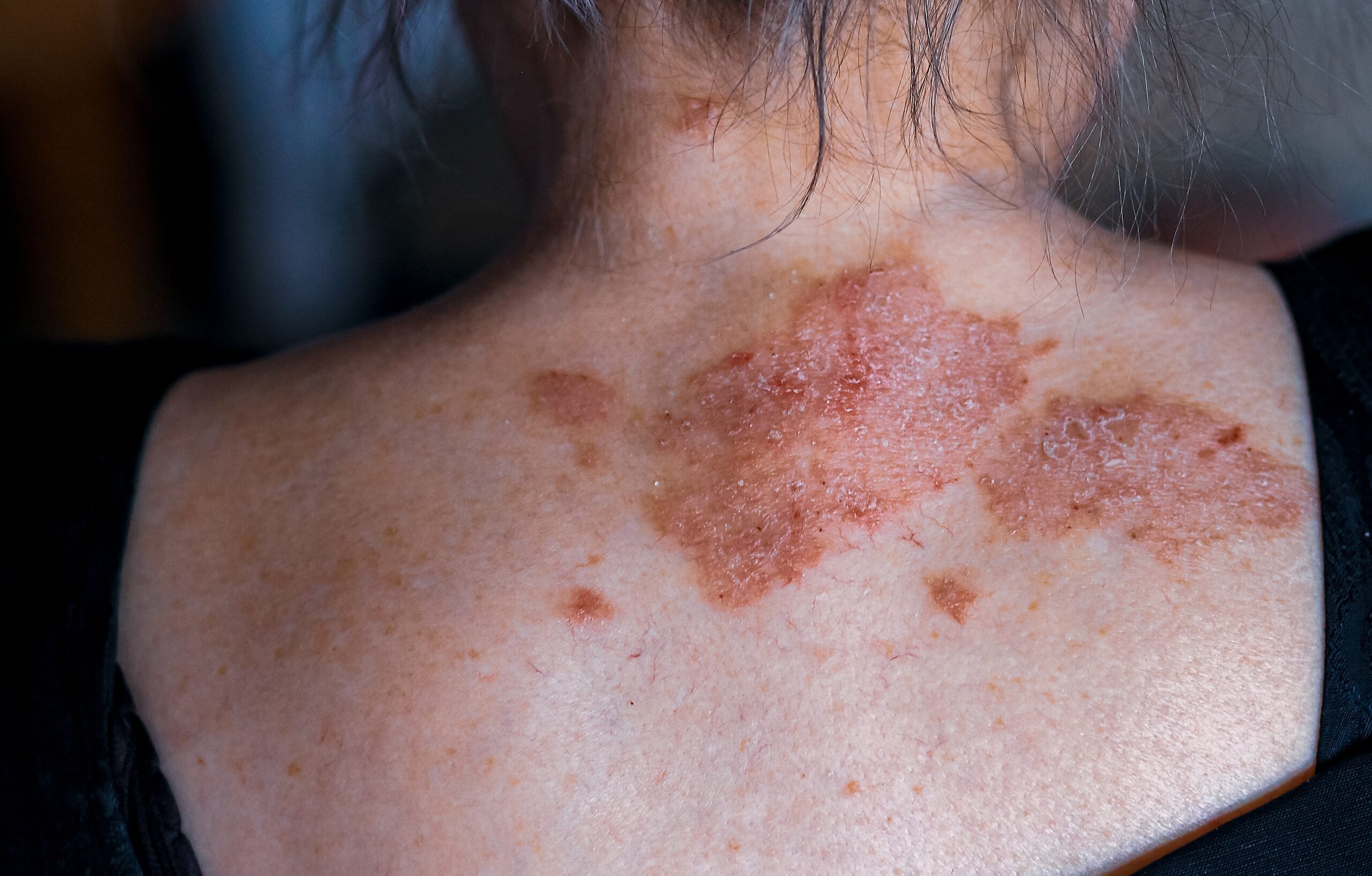
A café-au-lait macule is a common type of birthmark characterized by a hyperpigmented skin patch with a sharp border and a diameter larger than 0.5 cm. It is also called circumscribed café-au-lait hypermelanosis, von Recklinghausen spot, or simply ‘CALM’. These macules typically appear at birth (congenital) or during early infancy, though they can become noticeable later in infancy, especially after exposure to sunlight, which darkens the color.
Café-au-lait macules can occur in isolation or be associated with systemic diseases such as neurofibromatosis (NF), McCune Albright syndrome, Legius syndrome, and Noonan syndrome with multiple lentigines syndrome.
The prevalence of café-au-lait macules varies with race: 0.3% of Caucasians, 0.4% of Chinese, 3% of Hispanics, and 8% of African Americans.
Isolated café-au-lait macules are typically solitary. However, having more than three in Caucasians or more than five in African Americans is rare and should prompt further evaluation, referral, and close monitoring.
The brown color of café-au-lait macules results from the presence of melanin, a pigment produced by skin cells called melanocytes. In isolated café-au-lait macules, there is an excessive number of melanosomes (intracellular pigment granules) in the epidermal melanocytes, a condition known as epidermal melanotic hypermelanosis. In macules associated with NF type 1 and Leopard syndrome, there is an increased proliferation of epidermal melanocytes (epidermal melanocytic hyperplasia).
It is important to note that a café-au-lait macule is not classified as a congenital melanocytic nevus (a type of birthmark that can become cancerous).
Multiple café-au-lait macules may be related to various genetic syndromes. For instance, about half of those with neurofibromatosis type 1 (NF1) have an inherited mutation of the NF1 gene, while others may have sporadic mutations of the same gene.
Several other syndromes, such as NF type 2, Legius syndrome, McCune Albright syndrome, Noonan syndrome with multiple lentigines, Watson syndrome, Bloom syndrome, and Silver-Russell syndrome, can also present with café-au-lait macules. These syndromes have additional characteristic features and may require genetic testing for confirmation.
Diagnosing café-au-lait macules is usually done through clinical examination. A thorough clinical evaluation is needed to identify potentially associated syndromes if numerous or large macules are present. Syndromes can be diagnosed based on clinical manifestations or through genetic testing.
In terms of treatment, café-au-lait macules do not require medical intervention. Some cases have been treated with lasers, such as pulsed-dye laser, Er:YAG laser, Q-switched Nd:YAG laser, and Q-switched ruby or alexandrite laser. However, results from laser treatments have been inconsistent, and there are potential risks, including hyperpigmentation, hypopigmentation, and scarring.
Without treatment, café-au-lait macules typically persist throughout a person’s life. Recurrence rates are low for those who respond to laser treatment, but the outcomes are not consistent for all cases. The treatment of associated syndromes may be complex and require multidisciplinary care.


