Basal Cell Carcinoma (BCC), a common keratinocyte cancer also referred to as nonmelanoma cancer, is the most prevalent form of skin cancer. Known by names such as rodent ulcer and basalioma, individuals with BCC frequently experience multiple primary tumors over time.
Several factors increase the risk of developing BCC. It is particularly common in elderly males, though it also affects females and younger adults. Other risk factors include having had a previous BCC or other skin cancers like squamous cell carcinoma and melanoma; sun damage including photoaging and actinic keratoses, and a history of repeated sunburns. People with fair skin, blue eyes, and blond or red hair are more susceptible, although BCC can affect darker skin types as well. Previous skin injuries, thermal burns, diseases like cutaneous lupus and sebaceous naevus, and inherited syndromes such as basal cell naevus syndrome (Gorlin syndrome), Bazex-Dupré-Christol syndrome, Rombo syndrome, Oley syndrome, and xeroderma pigmentosum also heighten the risk. Additional factors include ionizing radiation, exposure to arsenic, immune suppression from disease or medications, and the use of certain medicines like hydrochlorothiazide.
BCC arises from multifactorial causes, predominantly DNA mutations in the patched (PTCH) tumor suppressor gene, part of the hedgehog signalling pathway, often triggered by ultraviolet radiation exposure. Both spontaneous and inherited gene defects can predispose individuals to BCC.
Characteristically, BCC is a locally invasive skin tumor exhibiting a slowly growing plaque or nodule, which may be skin colored, pink, or pigmented, and vary in size. It may spontaneously bleed or ulcerate. While BCC rarely poses a life threat, a small proportion can grow rapidly, invade deeply, or metastasize to local lymph nodes.
BCC manifests in several clinical types and over 20 histological growth patterns.
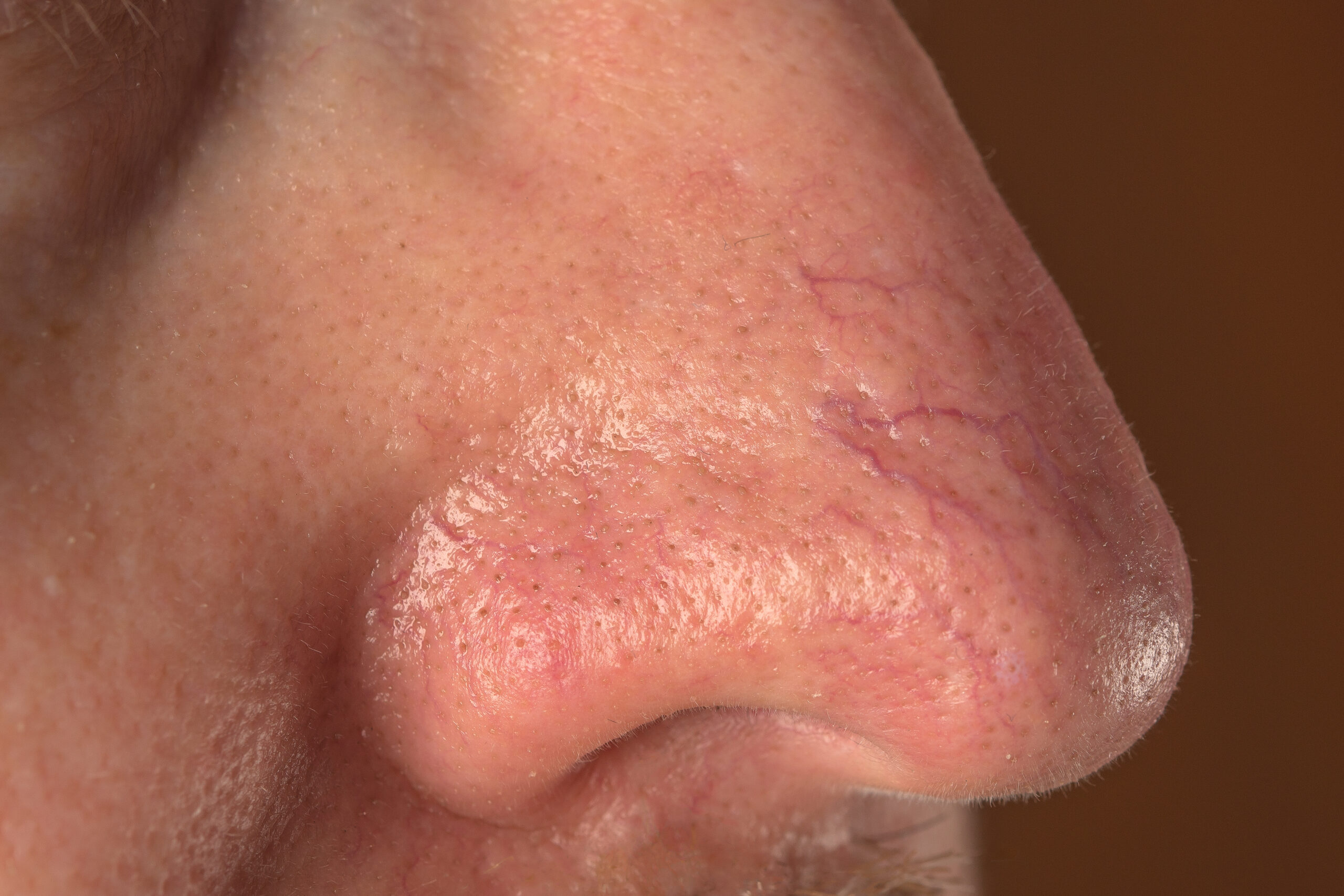
Nodular BCC, the most common type on the face, appears as a shiny or pearly nodule with a smooth surface, potentially with a central depression or ulceration, giving it a rolled edge appearance, and blood vessels visible across its surface. The cystic variant is soft with jelly-like contents. Micronodular, microcystic, and infiltrative types are more aggressive subtypes known as nodulocystic carcinoma.
Superficial BCC, often found in younger adults and commonly located on the upper trunk and shoulders, presents as a slightly scaly, irregular plaque with a thin, translucent rolled border and multiple microerosions.
Morphoeic BCC, typically located in mid-facial areas, resembles a waxy, scar-like plaque with indistinct borders and can infiltrate cutaneous nerves (perineural spread). It is also known as morpheic, morphoeiform, or sclerosing BCC.
Basosquamous carcinoma, a mixture of basal cell carcinoma (BCC) and squamous cell carcinoma (SCC), exhibits an infiltrative growth pattern and can be more aggressive than other BCC forms.
BCC recurrence after initial treatment is not uncommon, particularly in cases of incomplete excision, narrow margins at primary excision, or when it involves morphoeic, micronodular, and infiltrative subtypes, especially if located on the head and neck.
Advanced BCCs are large, often neglected tumors that can be several centimetres in diameter, deeply infiltrating, and challenging or impossible to treat surgically.
Metastatic BCC is extremely rare, often arising from large, neglected, or recurrent primary tumors on the head and neck, with aggressive subtypes. These may have undergone multiple prior treatments and can be fatal.
BCC diagnosis is typically clinical, based on the presence of a characteristic, slowly enlarging skin lesion. The diagnosis and histological subtype are usually confirmed pathologically by a diagnostic biopsy or following excision. Some superficial BCCs on the trunk and limbs are clinically diagnosed and treated non-surgically without histology confirmation.


