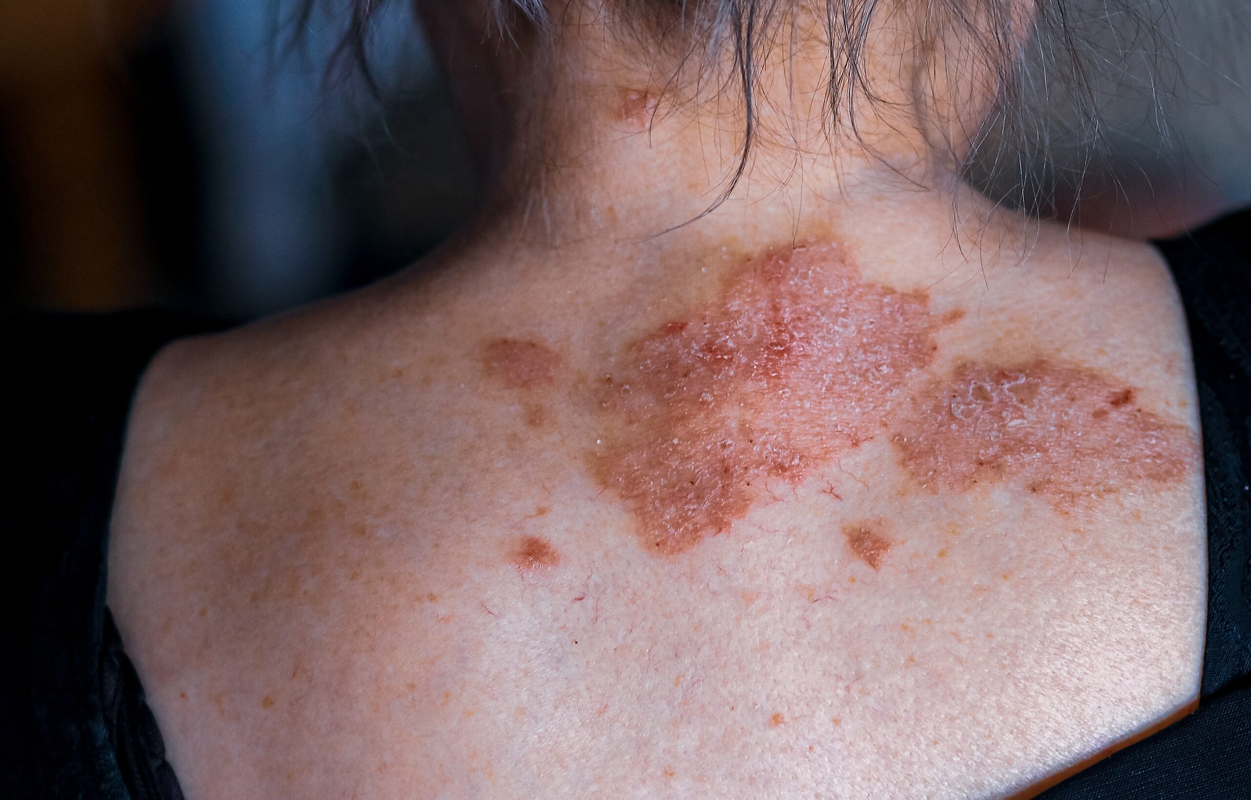
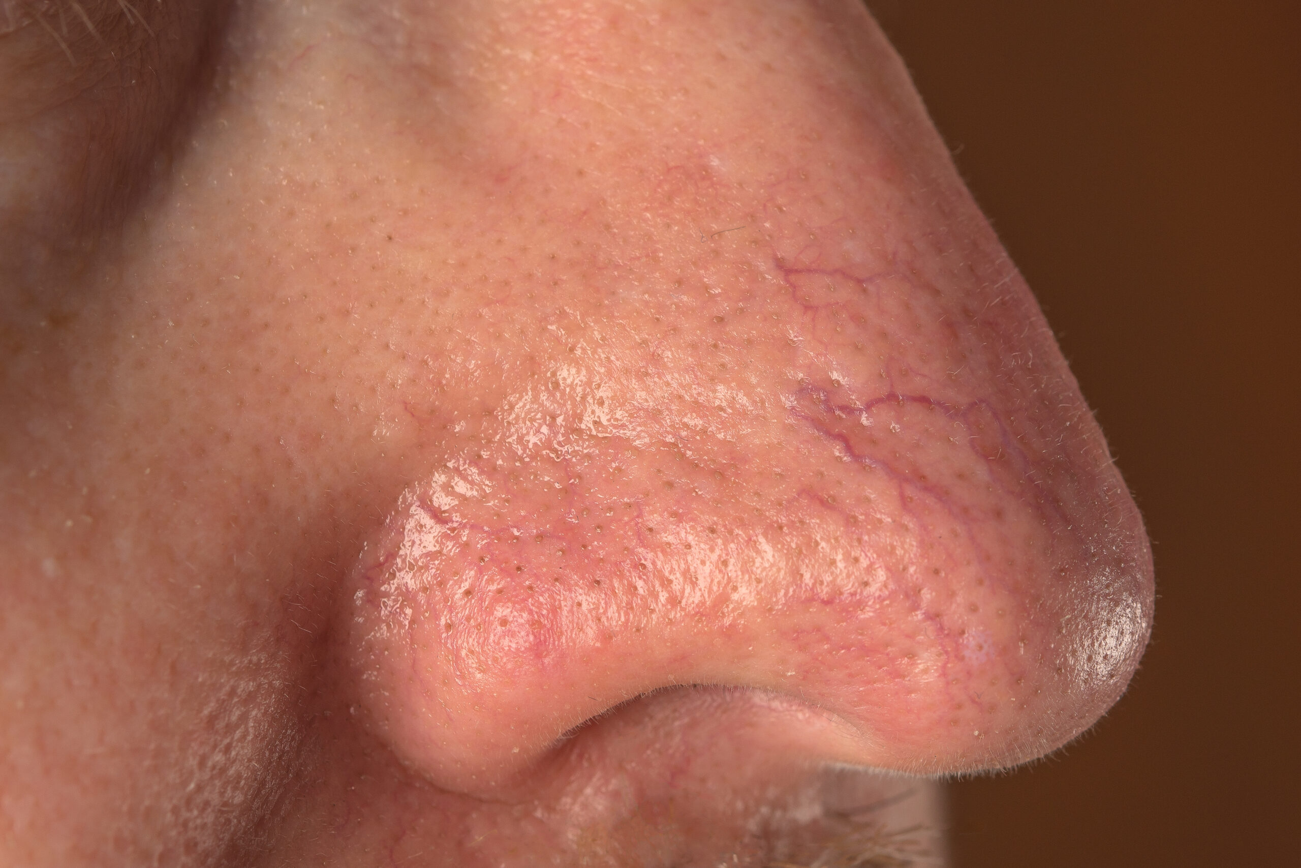
Acral lentiginous melanoma (ALM), a less common variant of melanoma, originates on the palms, soles, or beneath the nails and is alternatively known as acral melanoma. Initially, ALM may present as a dark brown or black macule exhibiting varied coloring, and in later stages, it may become nodular and ulcerated. This subtype grows slowly, potentially taking months to years before it becomes invasive, extending beyond the epidermis’s basement membrane where it starts.
Acral lentiginous melanoma accounts for only 2–3% of all melanoma diagnoses, making it the least frequently diagnosed subtype overall. However, it represents the most prevalent melanoma subtype among individuals with darker skin, comprising 40–60% of melanoma cases in Asian and African-American populations, whereas it is less common among Caucasians. The incidence of ALM tends to increase with age, with the median age at diagnosis being 63 years, affecting males and females equally, though females often receive a diagnosis earlier than males.
Typically, ALM emerges spontaneously, though there have been occasional reports of its development from pre-existing melanocytic naevi. The exact cause of ALM remains unclear. Unlike other melanoma subtypes, sunlight exposure is not considered a significant risk factor, although some case reports have suggested a UV signature similar to other melanomas. Theories on ALM’s origins include mechanical stress on the foot and genetic mutations, such as in the KIT, BRAF, NRAS, NF1, cyclin D1, CDKN2A, and MYC genes. The pathogenesis involves the uncontrolled proliferation of melanocytes, which produce pigment deep within the epidermis. Often, ALM is first noticed as a new pigmented macule varying from light to dark brown on the palm, sole, or under a nail, typically exhibiting a higher frequency on the feet due to a greater melanocyte density. In some cases, ALM appears as an amelanotic or hypomelanotic lesion, challenging to distinguish from benign conditions like warts or calluses.
Subungual melanoma, originating from the nail matrix, typically manifests as a longitudinal brown or black band within the fingernail or toenail, sometimes accompanied by nail dystrophy. Hutchinson’s sign, the involvement of the proximal nail fold, is a diagnostic indicator.
Invasive melanoma may be suggested by features such as nodularity, dark pigmentation, ulceration, itchiness, an increase in color variety, especially blue and black, and pigment spreading beyond the nail fold.
Untreated ALM can lead to complications like distal metastasis, while surgical management might result in scarring, skin contractures, digit amputation, phantom limb pain, functional impairment, and loss of nails. Diagnosis primarily relies on clinical history and presentation, supported by dermoscopy, which may reveal a parallel ridge pattern, absence of acral naevi characteristics, blotches, asymmetry, multiple colors, and diffuse pigmentation or stripes on the nail. Histopathology following excision or biopsy confirms the diagnosis, with staging based on Breslow depth, ulceration, and sentinel lymph node status, showing asymmetrical melanocyte proliferation at the dermoepidermal junction.
ALM’s differential diagnosis is wide-ranging, including acral naevi, trauma-related hemorrhage, calcaneal petechiae, tinea nigra, and soil stains, with pigmented actinic keratoses and seborrheic keratoses unlikely on acral surfaces. Amelanotic or hypomelanotic lesions may resemble benign conditions like warts or calluses.
Treatment focuses on wide local excision, with further actions determined by the Breslow thickness. Excision margins vary based on in-situ presence and Breslow depth, potentially necessitating skin grafts, flaps, or partial digit amputation. Additional treatments might include radiotherapy or immunotherapy.
Preventive measures for ALM are not well-established, but regular skin examinations and general sun protection are advised. Early medical review of new lesions on the palms, soles, or under nails is crucial. Compared to other melanomas, ALM typically has a poorer prognosis due to later-stage diagnosis and possibly genetic and biological differences influencing tumor aggression. Prognosis depends on factors like Breslow thickness, staging, and sentinel lymph node involvement, with age and gender also playing roles.


